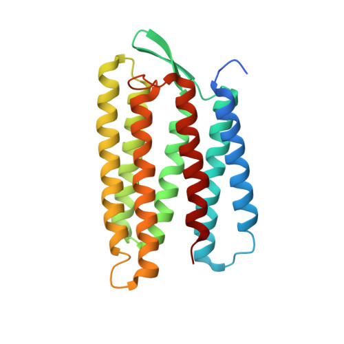
Proton uptake mechanism in bacteriorhodopsin captured by serial synchrotron crystallography.
Weinert, T., Skopintsev, P., James, D., Dworkowski, F., Panepucci, E., Kekilli, D., Furrer, A., Brunle, S., Mous, S., Ozerov, D., Nogly, P., Wang, M., Standfuss, J.(2019) Science 365: 61-65
- PubMed: 31273117
- DOI: https://doi.org/10.1126/science.aaw8634
- Primary Citation of Related Structures:
6RNJ, 6RPH, 6RQO, 6RQP - PubMed Abstract:
Conformational dynamics are essential for proteins to function. We adapted time-resolved serial crystallography developed at x-ray lasers to visualize protein motions using synchrotrons. We recorded the structural changes in the light-driven proton-pump bacteriorhodopsin over 200 milliseconds in time. The snapshot from the first 5 milliseconds after photoactivation shows structural changes associated with proton release at a quality comparable to that of previous x-ray laser experiments. From 10 to 15 milliseconds onwards, we observe large additional structural rearrangements up to 9 angstroms on the cytoplasmic side. Rotation of leucine-93 and phenylalanine-219 opens a hydrophobic barrier, leading to the formation of a water chain connecting the intracellular aspartic acid-96 with the retinal Schiff base. The formation of this proton wire recharges the membrane pump with a proton for the next cycle.
Organizational Affiliation:
Division of Biology and Chemistry-Laboratory for Biomolecular Research, Paul Scherrer Institut, 5232 Villigen, Switzerland. [email protected].


