6SLT

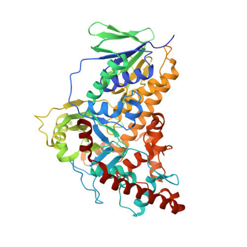
Flavin-dependent tryptophan 6-halogenase Thal in complex with tryptophan and FAD


Experimental Data Snapshot

  • Method: X-RAY DIFFRACTION
  • Resolution: 2.70 Å
  • R-Value Free: 0.244 
  • R-Value Work: 0.186 
  • R-Value Observed: 0.189 

Starting Model: experimental
View more details

wwPDB Validation   3D Report Full Report


Ligand Structure Quality Assessment 


This is version 1.3 of the entry. See complete history


Literature

Binding of FAD and tryptophan to the tryptophan 6-halogenase Thal is negatively coupled.

Moritzer, A.C.Niemann, H.H.

(2019) Protein Sci 28: 2112-2118

  • DOI: https://doi.org/10.1002/pro.3739
  • Primary Citation of Related Structures:  
    6SLS, 6SLT

  • PubMed Abstract: 

    Flavin-dependent halogenases require reduced flavin adenine dinucleotide (FADH 2 ), O 2 , and halide salts to halogenate their substrates. We describe the crystal structures of the tryptophan 6-halogenase Thal in complex with FAD or with both tryptophan and FAD. If tryptophan and FAD were soaked simultaneously, both ligands showed impaired binding and in some cases only the adenosine monophosphate or the adenosine moiety of FAD was resolved, suggesting that tryptophan binding increases the mobility mainly of the flavin mononucleotide moiety. This confirms a negative cooperativity between the binding of substrate and cofactor that was previously described for other tryptophan halogenases. Binding of substrate to tryptophan halogenases reduces the affinity for the oxidized cofactor FAD presumably to facilitate the regeneration of FADH 2 by flavin reductases.


  • Organizational Affiliation

    Department of Chemistry, Bielefeld University, Bielefeld, Germany.


Macromolecules
Find similar proteins by:  (by identity cutoff)  |  3D Structure
Entity ID: 1
MoleculeChains Sequence LengthOrganismDetailsImage
Tryptophan 6-halogenase
A, B
534Streptomyces albogriseolusMutation(s): 0 
Gene Names: thalthdH
EC: 1.14.19.59
UniProt
Find proteins for A1E280 (Streptomyces albogriseolus)
Explore A1E280 
Go to UniProtKB:  A1E280
Entity Groups  
Sequence Clusters30% Identity50% Identity70% Identity90% Identity95% Identity100% Identity
UniProt GroupA1E280
Sequence Annotations
Expand
  • Reference Sequence
Small Molecules
Ligands 5 Unique
IDChains Name / Formula / InChI Key2D Diagram3D Interactions
FAD (Subject of Investigation/LOI)
Query on FAD

Download Ideal Coordinates CCD File 
N [auth B]FLAVIN-ADENINE DINUCLEOTIDE
C27 H33 N9 O15 P2
VWWQXMAJTJZDQX-UYBVJOGSSA-N
AMP
Query on AMP

Download Ideal Coordinates CCD File 
K [auth A]ADENOSINE MONOPHOSPHATE
C10 H14 N5 O7 P
UDMBCSSLTHHNCD-KQYNXXCUSA-N
TRP
Query on TRP

Download Ideal Coordinates CCD File 
J [auth A]TRYPTOPHAN
C11 H12 N2 O2
QIVBCDIJIAJPQS-VIFPVBQESA-N
PO4
Query on PO4

Download Ideal Coordinates CCD File 
H [auth A],
I [auth A],
M [auth B]
PHOSPHATE ION
O4 P
NBIIXXVUZAFLBC-UHFFFAOYSA-K
GOL
Query on GOL

Download Ideal Coordinates CCD File 
C [auth A]
D [auth A]
E [auth A]
F [auth A]
G [auth A]
C [auth A],
D [auth A],
E [auth A],
F [auth A],
G [auth A],
L [auth B]
GLYCEROL
C3 H8 O3
PEDCQBHIVMGVHV-UHFFFAOYSA-N
Experimental Data & Validation

Experimental Data

  • Method: X-RAY DIFFRACTION
  • Resolution: 2.70 Å
  • R-Value Free: 0.244 
  • R-Value Work: 0.186 
  • R-Value Observed: 0.189 
  • Space Group: P 64
Unit Cell:
Length ( Å )Angle ( ˚ )
a = 138.25α = 90
b = 138.25β = 90
c = 142.01γ = 120
Software Package:
Software NamePurpose
REFMACrefinement
PDB_EXTRACTdata extraction
XDSdata reduction
XSCALEdata scaling
PHASERphasing

Structure Validation

View Full Validation Report


Ligand Structure Quality Assessment 


Entry History 

Deposition Data

Revision History  (Full details and data files)

  • Version 1.0: 2019-10-16
    Type: Initial release
  • Version 1.1: 2019-10-30
    Changes: Data collection, Database references
  • Version 1.2: 2019-11-27
    Changes: Database references
  • Version 1.3: 2024-01-24
    Changes: Data collection, Database references, Refinement description