6STZ

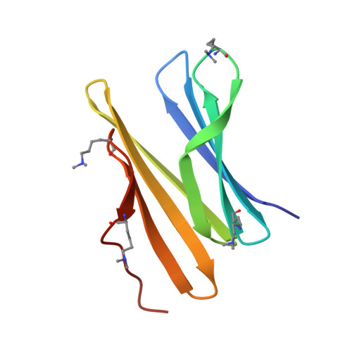
Crystal structure of dimethylated RSLex - cucurbituril free form


Experimental Data Snapshot

  • Method: X-RAY DIFFRACTION
  • Resolution: 1.14 Å
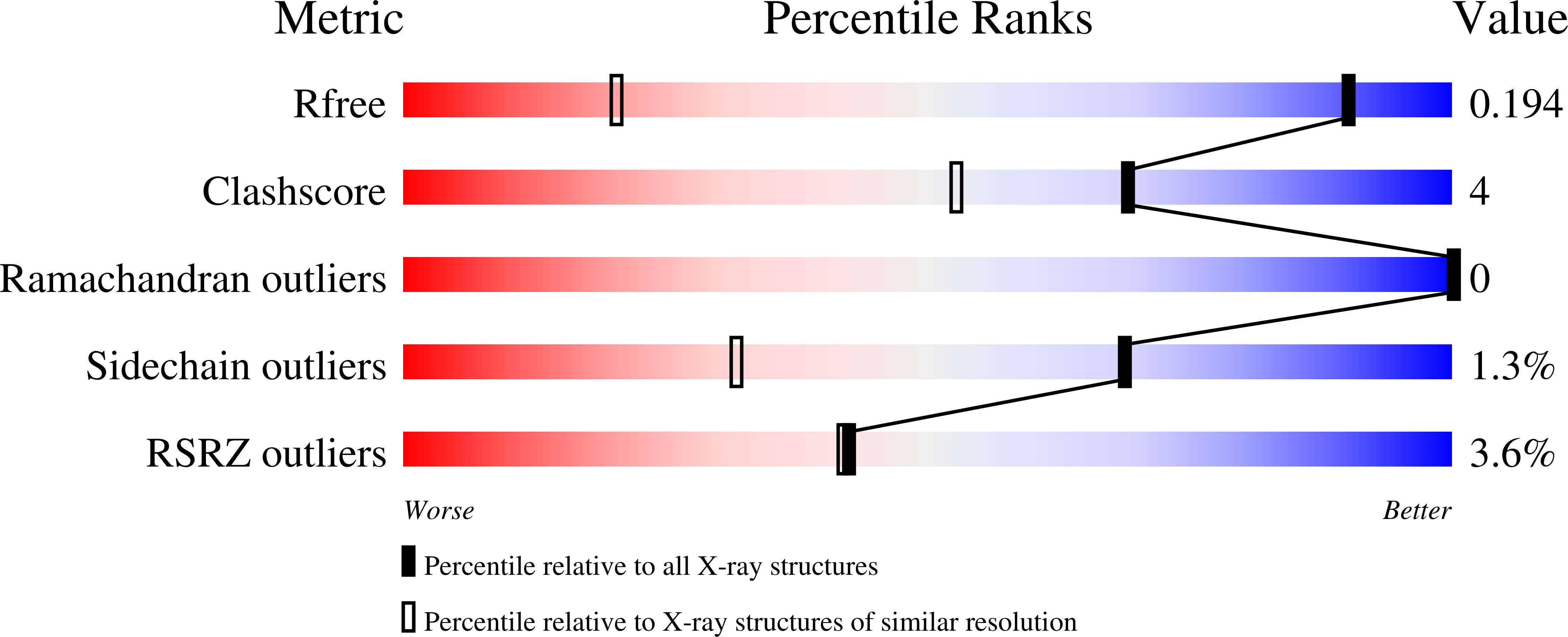
  • R-Value Free: 0.198 
  • R-Value Work: 0.173 
  • R-Value Observed: 0.174 

Starting Model: experimental
View more details

wwPDB Validation   3D Report Full Report


Ligand Structure Quality Assessment 


This is version 1.2 of the entry. See complete history


Literature

Engineered assembly of a protein-cucurbituril biohybrid.

Guagnini, F.Engilberge, S.Ramberg, K.O.Perez, J.Crowley, P.B.

(2020) Chem Commun (Camb) 56: 360-363

  • DOI: https://doi.org/10.1039/c9cc07198a
  • Primary Citation of Related Structures:  
    6STZ, 6SU0

  • PubMed Abstract: 

    A crystalline biohybrid with a 4 : 1 protein : cucurbituril mass ratio is presented. This result was achieved by engineering additional cucurbit[7]uril (Q7) binding sites into a β-propeller protein. In contrast to the parent protein, Q7-controlled assembly of the engineered variant occurred in solution, as evidenced by NMR and SAXS measurements.


  • Organizational Affiliation

    School of Chemistry, National University of Ireland Galway, University Road, Galway, H91 TK33, Ireland. [email protected].


Macromolecules
Find similar proteins by:  (by identity cutoff)  |  3D Structure
Entity ID: 1
MoleculeChains Sequence LengthOrganismDetailsImage
Fucose-binding lectin protein
A, B, C, D, E
A, B, C, D, E, F
89Ralstonia solanacearumMutation(s): 1 
Gene Names: rslRUN215_v1_180133
UniProt
Find proteins for Q8XXK6 (Ralstonia nicotianae (strain ATCC BAA-1114 / GMI1000))
Explore Q8XXK6 
Go to UniProtKB:  Q8XXK6
Entity Groups  
Sequence Clusters30% Identity50% Identity70% Identity90% Identity95% Identity100% Identity
UniProt GroupQ8XXK6
Sequence Annotations
Expand
  • Reference Sequence
Small Molecules
Ligands 1 Unique
IDChains Name / Formula / InChI Key2D Diagram3D Interactions
BDF
Query on BDF

Download Ideal Coordinates CCD File 
G [auth A]
H [auth A]
I [auth B]
J [auth B]
K [auth C]
G [auth A],
H [auth A],
I [auth B],
J [auth B],
K [auth C],
L [auth C],
M [auth D],
N [auth D],
O [auth E],
P [auth E],
Q [auth F],
R [auth F]
beta-D-fructopyranose
C6 H12 O6
LKDRXBCSQODPBY-ARQDHWQXSA-N
Modified Residues  1 Unique
IDChains TypeFormula2D DiagramParent
MLY
Query on MLY
A, B, C, D, E
A, B, C, D, E, F
L-PEPTIDE LINKINGC8 H18 N2 O2LYS
Experimental Data & Validation

Experimental Data

  • Method: X-RAY DIFFRACTION
  • Resolution: 1.14 Å
  • R-Value Free: 0.198 
  • R-Value Work: 0.173 
  • R-Value Observed: 0.174 
  • Space Group: P 32
Unit Cell:
Length ( Å )Angle ( ˚ )
a = 43.948α = 90
b = 43.948β = 90
c = 207.273γ = 120
Software Package:
Software NamePurpose
BUSTERrefinement
Aimlessdata scaling
PDB_EXTRACTdata extraction
XDSdata reduction
PHASERphasing

Structure Validation

View Full Validation Report


Ligand Structure Quality Assessment 


Entry History & Funding Information

Deposition Data


Funding OrganizationLocationGrant Number
Science Foundation IrelandIreland13/CDA/2168

Revision History  (Full details and data files)

  • Version 1.0: 2020-02-19
    Type: Initial release
  • Version 1.1: 2020-07-29
    Type: Remediation
    Reason: Carbohydrate remediation
    Changes: Data collection, Derived calculations, Structure summary
  • Version 1.2: 2024-01-24
    Changes: Data collection, Database references, Refinement description, Structure summary