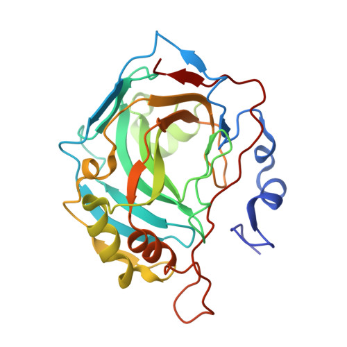
6T4O

Human Carbonic anhydrase II bound by 3,5-dimethylbenzenesulfonamide


Experimental Data Snapshot

  • Method: X-RAY DIFFRACTION
  • Resolution: 1.13 Å
  • R-Value Free: 0.166 
  • R-Value Work: 0.139 
  • R-Value Observed: 0.142 

Starting Model: experimental
View more details

wwPDB Validation   3D Report Full Report


Ligand Structure Quality Assessment 


This is version 1.2 of the entry. See complete history


Literature

Isoform-Selective Enzyme Inhibitors by Exploring Pocket Size According to the Lock-and-Key Principle.

Dudutiene, V.Zubriene, A.Kairys, V.Smirnov, A.Smirnoviene, J.Leitans, J.Kazaks, A.Tars, K.Manakova, L.Grazulis, S.Matulis, D.

(2020) Biophys J 119: 1513-1524

  • DOI: https://doi.org/10.1016/j.bpj.2020.08.037
  • Primary Citation of Related Structures:  
    6T4N, 6T4O, 6T4P, 6T5C, 6T5P, 6T5Q, 6T81, 6TL5, 6TL6

  • PubMed Abstract: 

    In the design of high-affinity and enzyme isoform-selective inhibitors, we applied an approach of augmenting the substituents attached to the benzenesulfonamide scaffold in three ways, namely, substitutions at the 3,5- or 2,4,6-positions or expansion of the condensed ring system. The increased size of the substituents determined the spatial limitations of the active sites of the 12 catalytically active human carbonic anhydrase (CA) isoforms until no binding was observed because of the inability of the compounds to fit in the active site. This approach led to the discovery of high-affinity and high-selectivity compounds for the anticancer target CA IX and antiobesity target CA VB. The x-ray crystallographic structures of compounds bound to CA IX showed the positions of the bound compounds, whereas computational modeling confirmed that steric clashes prevent the binding of these compounds to other isoforms and thus avoid undesired side effects. Such an approach, based on the Lock-and-Key principle, could be used for the development of enzyme-specific drug candidate compounds.


  • Organizational Affiliation

    Department of Biothermodynamics and Drug Design, Vilnius University, Vilnius, Lithuania.


Macromolecules
Find similar proteins by:  (by identity cutoff)  |  3D Structure
Entity ID: 1
MoleculeChains Sequence LengthOrganismDetailsImage
carbonic anhydrase 2260Homo sapiensMutation(s): 0 
EC: 4.2.1.1 (PDB Primary Data), 4.2.1.69 (UniProt)
UniProt & NIH Common Fund Data Resources
Find proteins for P00918 (Homo sapiens)
Explore P00918 
Go to UniProtKB:  P00918
PHAROS:  P00918
GTEx:  ENSG00000104267 
Entity Groups  
Sequence Clusters30% Identity50% Identity70% Identity90% Identity95% Identity100% Identity
UniProt GroupP00918
Sequence Annotations
Expand
  • Reference Sequence
Experimental Data & Validation

Experimental Data

  • Method: X-RAY DIFFRACTION
  • Resolution: 1.13 Å
  • R-Value Free: 0.166 
  • R-Value Work: 0.139 
  • R-Value Observed: 0.142 
  • Space Group: P 1 21 1
Unit Cell:
Length ( Å )Angle ( ˚ )
a = 42.226α = 90
b = 41.325β = 104.31
c = 72.049γ = 90
Software Package:
Software NamePurpose
SCALAdata scaling
REFMACrefinement
PDB_EXTRACTdata extraction
XDSdata reduction
MOLREPphasing
Cootmodel building

Structure Validation

View Full Validation Report


Ligand Structure Quality Assessment 


Entry History 

Deposition Data

Revision History  (Full details and data files)

  • Version 1.0: 2020-10-14
    Type: Initial release
  • Version 1.1: 2020-10-28
    Changes: Database references
  • Version 1.2: 2024-01-24
    Changes: Data collection, Database references, Refinement description