6T9A

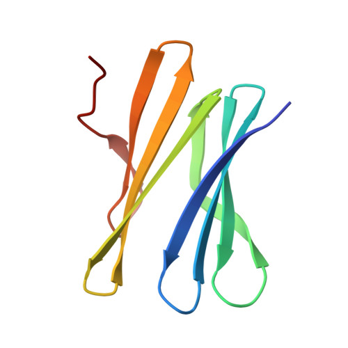
Crystal structrue of RSL W31FW76F lectin mutant in complex with L-fucose


Experimental Data Snapshot

  • Method: X-RAY DIFFRACTION
  • Resolution: 2.00 Å
  • R-Value Free: 0.198 
  • R-Value Work: 0.152 
  • R-Value Observed: 0.155 

Starting Model: experimental
View more details

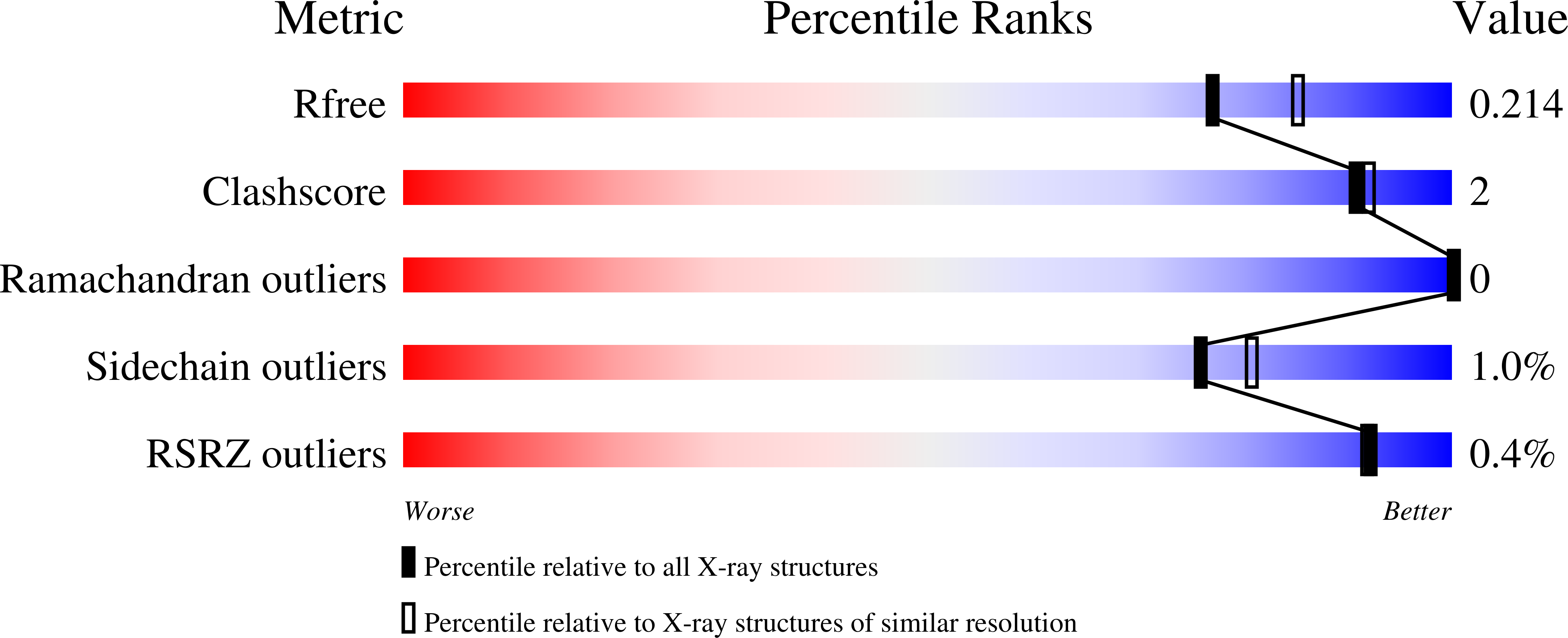
wwPDB Validation   3D Report Full Report


Ligand Structure Quality Assessment 


This is version 1.4 of the entry. See complete history


Literature

The CH-pi Interaction in Protein-Carbohydrate Binding: Bioinformatics and In Vitro Quantification.

Houser, J.Kozmon, S.Mishra, D.Hammerova, Z.Wimmerova, M.Koca, J.

(2020) Chemistry 26: 10769-10780

  • DOI: https://doi.org/10.1002/chem.202000593
  • Primary Citation of Related Structures:  
    6T99, 6T9A, 6T9B

  • PubMed Abstract: 

    The molecular recognition of carbohydrates by proteins plays a key role in many biological processes including immune response, pathogen entry into a cell, and cell-cell adhesion (e.g., in cancer metastasis). Carbohydrates interact with proteins mainly through hydrogen bonding, metal-ion-mediated interaction, and non-polar dispersion interactions. The role of dispersion-driven CH-π interactions (stacking) in protein-carbohydrate recognition has been underestimated for a long time considering the polar interactions to be the main forces for saccharide interactions. However, over the last few years it turns out that non-polar interactions are equally important. In this study, we analyzed the CH-π interactions employing bioinformatics (data mining, structural analysis), several experimental (isothermal titration calorimetry (ITC), X-ray crystallography), and computational techniques. The Protein Data Bank (PDB) has been used as a source of structural data. The PDB contains over 12 000 protein complexes with carbohydrates. Stacking interactions are very frequently present in such complexes (about 39 % of identified structures). The calculations and the ITC measurement results suggest that the CH-π stacking contribution to the overall binding energy ranges from 4 up to 8 kcal mol -1 . All the results show that the stacking CH-π interactions in protein-carbohydrate complexes can be considered to be a driving force of the binding in such complexes.


  • Organizational Affiliation

    Central European Institute of Technology, Masaryk University, Kamenice 5, 62500, Brno, Czech Republic.


Macromolecules
Find similar proteins by:  (by identity cutoff)  |  3D Structure
Entity ID: 1
MoleculeChains Sequence LengthOrganismDetailsImage
Fucose-binding lectin protein
A, B, C
90Ralstonia solanacearumMutation(s): 2 
Gene Names: RSP795_21825RSP799_05830RSP822_19650RUN39_v1_50103
UniProt
Find proteins for A0A0S4TLR1 (Ralstonia solanacearum)
Explore A0A0S4TLR1 
Go to UniProtKB:  A0A0S4TLR1
Entity Groups  
Sequence Clusters30% Identity50% Identity70% Identity90% Identity95% Identity100% Identity
UniProt GroupA0A0S4TLR1
Sequence Annotations
Expand
  • Reference Sequence
Small Molecules
Ligands 3 Unique
IDChains Name / Formula / InChI Key2D Diagram3D Interactions
FUL (Subject of Investigation/LOI)
Query on FUL

Download Ideal Coordinates CCD File 
E [auth A]
G [auth A]
J [auth B]
K [auth B]
N [auth C]
E [auth A],
G [auth A],
J [auth B],
K [auth B],
N [auth C],
Q [auth C]
beta-L-fucopyranose
C6 H12 O5
SHZGCJCMOBCMKK-KGJVWPDLSA-N
FUC (Subject of Investigation/LOI)
Query on FUC

Download Ideal Coordinates CCD File 
D [auth A],
F [auth A],
I [auth B],
M [auth C],
P [auth C]
alpha-L-fucopyranose
C6 H12 O5
SHZGCJCMOBCMKK-SXUWKVJYSA-N
EDO
Query on EDO

Download Ideal Coordinates CCD File 
H [auth A],
L [auth B],
O [auth C]
1,2-ETHANEDIOL
C2 H6 O2
LYCAIKOWRPUZTN-UHFFFAOYSA-N
Experimental Data & Validation

Experimental Data

  • Method: X-RAY DIFFRACTION
  • Resolution: 2.00 Å
  • R-Value Free: 0.198 
  • R-Value Work: 0.152 
  • R-Value Observed: 0.155 
  • Space Group: P 1
Unit Cell:
Length ( Å )Angle ( ˚ )
a = 40.262α = 116.4
b = 42.105β = 94.54
c = 44.02γ = 115.49
Software Package:
Software NamePurpose
SCALAdata scaling
REFMACrefinement
PDB_EXTRACTdata extraction
XDSdata reduction
MOLREPphasing

Structure Validation

View Full Validation Report


Ligand Structure Quality Assessment 


Entry History 

Deposition Data

Revision History  (Full details and data files)

  • Version 1.0: 2020-04-22
    Type: Initial release
  • Version 1.1: 2020-07-29
    Type: Remediation
    Reason: Carbohydrate remediation
    Changes: Data collection, Derived calculations, Structure summary
  • Version 1.2: 2020-08-12
    Changes: Database references, Structure summary
  • Version 1.3: 2020-12-16
    Changes: Database references
  • Version 1.4: 2024-01-24
    Changes: Data collection, Database references, Refinement description