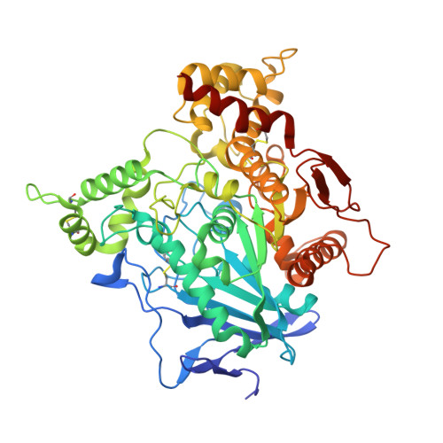
6U3P

Binary complex of native hAChE with oxime reactivator LG-703


Experimental Data Snapshot

  • Method: X-RAY DIFFRACTION
  • Resolution: 3.00 Å
  • R-Value Free: 0.253 
  • R-Value Work: 0.205 
  • R-Value Observed: 0.208 

Starting Model: experimental
View more details

wwPDB Validation   3D Report Full Report


Ligand Structure Quality Assessment 


This is version 1.4 of the entry. See complete history


Literature

Rational design, synthesis, and evaluation of uncharged, "smart" bis-oxime antidotes of organophosphate-inhibited human acetylcholinesterase.

Gorecki, L.Gerlits, O.Kong, X.Cheng, X.Blumenthal, D.K.Taylor, P.Ballatore, C.Kovalevsky, A.Radic, Z.

(2020) J Biol Chem 295: 4079-4092

  • DOI: https://doi.org/10.1074/jbc.RA119.012400
  • Primary Citation of Related Structures:  
    6U34, 6U37, 6U3P

  • PubMed Abstract: 

    Organophosphate (OP) intoxications from nerve agent and OP pesticide exposures are managed with pyridinium aldoxime-based therapies whose success rates are currently limited. The pyridinium cation hampers uptake of OPs into the central nervous system (CNS). Furthermore, it frequently binds to aromatic residues of OP-inhibited acetylcholinesterase (AChE) in orientations that are nonproductive for AChE reactivation, and the structural diversity of OPs impedes efficient reactivation. Improvements of OP antidotes need to include much better access of AChE reactivators to the CNS and optimized orientation of the antidotes' nucleophile within the AChE active-center gorge. On the basis of X-ray structures of a CNS-penetrating reactivator, monoxime RS194B, reversibly bound to native and venomous agent X (VX)-inhibited human AChE, here we created seven uncharged acetamido bis-oximes as candidate antidotes. Both oxime groups in these bis-oximes were attached to the same central, saturated heterocyclic core. Diverse protonation of the heterocyclic amines and oxime groups of the bis-oximes resulted in equilibration among up to 16 distinct ionization forms, including uncharged forms capable of diffusing into the CNS and multiple zwitterionic forms optimal for reactivation reactions. Conformationally diverse zwitterions that could act as structural antidote variants significantly improved in vitro reactivation of diverse OP-human AChE conjugates. Oxime group reorientation of one of the bis-oximes, forcing it to point into the active center for reactivation, was confirmed by X-ray structural analysis. Our findings provide detailed structure-activity properties of several CNS-directed, uncharged aliphatic bis-oximes holding promise for use as protonation-dependent, conformationally adaptive, "smart" accelerated antidotes against OP toxicity.


  • Organizational Affiliation

    Skaggs School of Pharmacy and Pharmaceutical Sciences, University of California San Diego, La Jolla, California 92093-0751.


Macromolecules
Find similar proteins by:  (by identity cutoff)  |  3D Structure
Entity ID: 1
MoleculeChains Sequence LengthOrganismDetailsImage
Acetylcholinesterase
A, B
550Homo sapiensMutation(s): 0 
Gene Names: ACHE
EC: 3.1.1.7
UniProt & NIH Common Fund Data Resources
Find proteins for P22303 (Homo sapiens)
Explore P22303 
Go to UniProtKB:  P22303
PHAROS:  P22303
GTEx:  ENSG00000087085 
Entity Groups  
Sequence Clusters30% Identity50% Identity70% Identity90% Identity95% Identity100% Identity
UniProt GroupP22303
Sequence Annotations
Expand
  • Reference Sequence
Small Molecules
Ligands 2 Unique
IDChains Name / Formula / InChI Key2D Diagram3D Interactions
PQY (Subject of Investigation/LOI)
Query on PQY

Download Ideal Coordinates CCD File 
C [auth A](2E,2'E)-N,N'-[1,4-diazepane-1,4-diyldi(ethane-2,1-diyl)]bis[2-(hydroxyimino)acetamide]
C13 H24 N6 O4
DFNMYOZHKNITMF-OTYYAQKOSA-N
DMS
Query on DMS

Download Ideal Coordinates CCD File 
D [auth B]DIMETHYL SULFOXIDE
C2 H6 O S
IAZDPXIOMUYVGZ-UHFFFAOYSA-N
Experimental Data & Validation

Experimental Data

  • Method: X-RAY DIFFRACTION
  • Resolution: 3.00 Å
  • R-Value Free: 0.253 
  • R-Value Work: 0.205 
  • R-Value Observed: 0.208 
  • Space Group: P 31
Unit Cell:
Length ( Å )Angle ( ˚ )
a = 125.407α = 90
b = 125.407β = 90
c = 129.826γ = 120
Software Package:
Software NamePurpose
PHENIXrefinement
HKL-3000data reduction
HKL-3000data scaling
PHASERphasing

Structure Validation

View Full Validation Report


Ligand Structure Quality Assessment 


Entry History & Funding Information

Deposition Data


Funding OrganizationLocationGrant Number
National Institutes of Health/National Institute of Neurological Disorders and Stroke (NIH/NINDS)United StatesU01 NS083451

Revision History  (Full details and data files)

  • Version 1.0: 2020-02-12
    Type: Initial release
  • Version 1.1: 2020-02-19
    Changes: Database references, Derived calculations
  • Version 1.2: 2020-04-08
    Changes: Database references
  • Version 1.3: 2023-10-11
    Changes: Data collection, Database references, Refinement description
  • Version 1.4: 2024-11-13
    Changes: Structure summary