Axial Heme Coordination by the Tyr-His Motif in the Extracellular Hemophore HasAp Is Critical for the Release of Heme to the HasR Receptor of Pseudomonas aeruginosa .
Dent, A.T., Brimberry, M., Albert, T., Lanzilotta, W.N., Moenne-Loccoz, P., Wilks, A.(2021) Biochemistry 60: 2549-2559
- PubMed: 34324310
- DOI: https://doi.org/10.1021/acs.biochem.1c00389
- Primary Citation of Related Structures:
6U87 - PubMed Abstract:
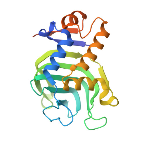
Pseudomonas aeruginosa senses extracellular heme via an extra cytoplasmic function σ factor that is activated upon interaction of the hemophore holo-HasAp with the HasR receptor. Herein, we show Y75H holo-HasAp interacts with HasR but is unable to release heme for signaling and uptake. To understand this inhibition, we undertook a spectroscopic characterization of Y75H holo-HasAp by resonance Raman (RR), electron paramagnetic resonance (EPR), and X-ray crystallography. The RR spectra are consistent with a mixed six-coordinate high-spin (6cHS), six-coordinate low-spin (6cLS) heme configuration and an H 2 18 O exchangeable Fe III -O stretching frequency with 16 O/ 18 O and H/D isotope shifts that support a two-body Fe-OH 2 oscillator with (iron-hydroxy)-like character as both hydrogen atoms are engaged in short hydrogen bond interactions with protein side chains. Further support comes from the EPR spectrum of Y75H holo-HasAp that shows a LS rhombic signal with ligand-field splitting values intermediate between those of His-hydroxy and bis-His ferric hemes. The crystal structure of Y75H holo-HasAp confirmed the coordinated solvent molecule hydrogen bonded through H75 and H83. The long-range conformational rearrangement of HasAp upon heme binding can still take place in Y75H holo-HasAp, because the intercalation of a hydroxy ligand between the heme iron and H75 allows the variant to reproduce the heme binding pocket observed in wild-type holo-HasAp. However, in the absence of a covalent linkage to the Y75 loop combined with the malleability provided by the bracketing H75 and H83 hydrogen bonds, either the hydroxy sixth ligand remains bound after complexation of Y75H holo-HasAp with HasR or rearrangement and coordination of H85 prevent heme transfer.
Organizational Affiliation:
Department of Pharmaceutical Sciences, School of Pharmacy, University of Maryland, 20 Penn Street, Baltimore, Maryland 21201, United States.


