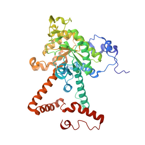
6WSI

Intact cis-2,3-epoxysuccinic acid bound to Isocitrate Lyase-1 from Mycobacterium tuberculosis


Experimental Data Snapshot

  • Method: X-RAY DIFFRACTION
  • Resolution: 1.75 Å
  • R-Value Free: 0.196 
  • R-Value Work: 0.164 
  • R-Value Observed: 0.165 

Starting Model: experimental
View more details

wwPDB Validation   3D Report Full Report


Ligand Structure Quality Assessment 


This is version 1.2 of the entry. See complete history


Literature

Covalent Inactivation of Mycobacterium tuberculosis Isocitrate Lyase by cis -2,3-Epoxy-Succinic Acid.

Pham, T.V.Mellott, D.M.Moghadamchargari, Z.Chen, K.Krieger, I.Laganowsky, A.Sacchettini, J.C.Meek, T.D.

(2021) ACS Chem Biol 16: 463-470

  • DOI: https://doi.org/10.1021/acschembio.0c00740
  • Primary Citation of Related Structures:  
    6VB9, 6WSI

  • PubMed Abstract: 

    The isocitrate lyases (ICL1/2) are essential enzymes of Mycobacterium tuberculosis ( Mtb ), the causative agent of tuberculosis. At present, no ICL1/2 inhibitors have progressed to clinical evaluation, despite extensive drug discovery efforts. Herein, we surveyed succinate analogs against ICL1 and found that dicarboxylic acids constrained in their synperiplanar conformations, such as maleic acid, comprise uncompetitive inhibitors of ICL1 and inhibit more potently than their trans -isomers. From this, we identified cis -2,3 epoxysuccinic acid ( cis -EpS) as a selective, irreversible covalent inactivator of Mtb ICL1 ( k inact / K inact = (5.0 ± 1.4) × 10 4 M -1 s -1 ; K inact = 200 ± 50 nM), the most potent inactivator of ICL1 yet characterized. Crystallographic and mass spectrometric analysis demonstrated that Cys 191 of ICL1 was S-malylated by cis -EpS, and a crystallographic "snapshot" of inactivation lent insight into the chemical mechanism of this inactivation. Proteomic analysis of E. coli lysates showed that cis -EpS selectively labeled plasmid-expressed Mtb ICL1. Consistently, cis -EpS, but not its trans -isomer, inhibited the growth of Mtb under conditions in which ICL function is essential. These findings encourage the development of analogs of cis -2,3-epoxysuccinate as antituberculosis agents.


  • Organizational Affiliation

    Department of Biochemistry and Biophysics, Texas A&M University, College Station, Texas 77843, United States.


Macromolecules
Find similar proteins by:  (by identity cutoff)  |  3D Structure
Entity ID: 1
MoleculeChains Sequence LengthOrganismDetailsImage
Isocitrate lyase
A, B, C, D
428Mycobacterium tuberculosisMutation(s): 0 
Gene Names: 
EC: 4.1.3.1
UniProt
Find proteins for P9WKK7 (Mycobacterium tuberculosis (strain ATCC 25618 / H37Rv))
Explore P9WKK7 
Go to UniProtKB:  P9WKK7
Entity Groups  
Sequence Clusters30% Identity50% Identity70% Identity90% Identity95% Identity100% Identity
UniProt GroupP9WKK7
Sequence Annotations
Expand
  • Reference Sequence
Small Molecules
Ligands 5 Unique
IDChains Name / Formula / InChI Key2D Diagram3D Interactions
U9S (Subject of Investigation/LOI)
Query on U9S

Download Ideal Coordinates CCD File 
GA [auth D],
H [auth A],
O [auth B],
Y [auth C]
(2R,3S)-oxirane-2,3-dicarboxylic acid
C4 H4 O5
DCEMCPAKSGRHCN-XIXRPRMCSA-N
PEG
Query on PEG

Download Ideal Coordinates CCD File 
CA [auth C],
K [auth A],
KA [auth D],
LA [auth D],
U [auth B]
DI(HYDROXYETHYL)ETHER
C4 H10 O3
MTHSVFCYNBDYFN-UHFFFAOYSA-N
GOL
Query on GOL

Download Ideal Coordinates CCD File 
AA [auth C]
BA [auth C]
HA [auth D]
I [auth A]
IA [auth D]
AA [auth C],
BA [auth C],
HA [auth D],
I [auth A],
IA [auth D],
J [auth A],
JA [auth D],
P [auth B],
Q [auth B],
R [auth B],
S [auth B],
T [auth B],
Z [auth C]
GLYCEROL
C3 H8 O3
PEDCQBHIVMGVHV-UHFFFAOYSA-N
GLV
Query on GLV

Download Ideal Coordinates CCD File 
FA [auth D],
G [auth A],
N [auth B],
X [auth C]
GLYOXYLIC ACID
C2 H2 O3
HHLFWLYXYJOTON-UHFFFAOYSA-N
MG
Query on MG

Download Ideal Coordinates CCD File 
DA [auth D]
E [auth A]
EA [auth D]
F [auth A]
L [auth B]
DA [auth D],
E [auth A],
EA [auth D],
F [auth A],
L [auth B],
M [auth B],
V [auth C],
W [auth C]
MAGNESIUM ION
Mg
JLVVSXFLKOJNIY-UHFFFAOYSA-N
Experimental Data & Validation

Experimental Data

  • Method: X-RAY DIFFRACTION
  • Resolution: 1.75 Å
  • R-Value Free: 0.196 
  • R-Value Work: 0.164 
  • R-Value Observed: 0.165 
  • Space Group: P 21 21 21
Unit Cell:
Length ( Å )Angle ( ˚ )
a = 79.17α = 90
b = 134.205β = 90
c = 161.036γ = 90
Software Package:
Software NamePurpose
PHENIXrefinement
XDSdata reduction
Aimlessdata scaling
PDB_EXTRACTdata extraction
MOLREPphasing

Structure Validation

View Full Validation Report


Ligand Structure Quality Assessment 


Entry History & Funding Information

Deposition Data


Funding OrganizationLocationGrant Number
National Institutes of Health/National Institute Of Allergy and Infectious Diseases (NIH/NIAID)United StatesP01A1095208

Revision History  (Full details and data files)

  • Version 1.0: 2021-03-24
    Type: Initial release
  • Version 1.1: 2021-03-31
    Changes: Database references
  • Version 1.2: 2023-10-18
    Changes: Data collection, Database references, Refinement description