7B5Y

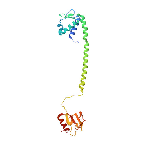
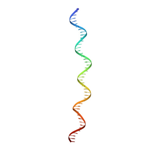
S. agalactiae BusR in complex with its busAB-promotor DNA


Experimental Data Snapshot

  • Method: ELECTRON MICROSCOPY
  • Resolution: 7.10 Å
  • Aggregation State: PARTICLE 
  • Reconstruction Method: SINGLE PARTICLE 

Starting Model: experimental
View more details

wwPDB Validation   3D Report Full Report


This is version 1.2 of the entry. See complete history


Literature

BusR senses bipartite DNA binding motifs by a unique molecular ruler architecture.

Bandera, A.M.Bartho, J.Lammens, K.Drexler, D.J.Kleinschwarzer, J.Hopfner, K.P.Witte, G.

(2021) Nucleic Acids Res 49: 10166-10177

  • DOI: https://doi.org/10.1093/nar/gkab736
  • Primary Citation of Related Structures:  
    7B5T, 7B5U, 7B5W, 7B5Y, 7OZ3

  • PubMed Abstract: 

    The cyclic dinucleotide second messenger c-di-AMP is a major player in regulation of potassium homeostasis and osmolyte transport in a variety of bacteria. Along with various direct interactions with proteins such as potassium channels, the second messenger also specifically binds to transcription factors, thereby altering the processes in the cell on the transcriptional level. We here describe the structural and biochemical characterization of BusR from the human pathogen Streptococcus agalactiae. BusR is a member of a yet structurally uncharacterized subfamily of the GntR family of transcription factors that downregulates transcription of the genes for the BusA (OpuA) glycine-betaine transporter upon c-di-AMP binding. We report crystal structures of full-length BusR, its apo and c-di-AMP bound effector domain, as well as cryo-EM structures of BusR bound to its operator DNA. Our structural data, supported by biochemical and biophysical data, reveal that BusR utilizes a unique domain assembly with a tetrameric coiled-coil in between the binding platforms, serving as a molecular ruler to specifically recognize a 22 bp separated bipartite binding motif. Binding of c-di-AMP to BusR induces a shift in equilibrium from an inactivated towards an activated state that allows BusR to bind the target DNA, leading to transcriptional repression.


  • Organizational Affiliation

    Gene Center, Ludwig-Maximilians-Universität München, Feodor-Lynen-Str. 25, D-81377 München, Germany.


Macromolecules

Find similar proteins by:  (by identity cutoff)  |  3D Structure
Entity ID: 1
MoleculeChains Sequence LengthOrganismDetailsImage
GntR family transcriptional regulatorA [auth C],
B [auth A],
C [auth B],
D
215Streptococcus agalactiaeMutation(s): 0 
Gene Names: BM110_ORF1201AX245_01365C6N10_09995F5043_05515GD434_05225RDF_1124
UniProt
Find proteins for K0JNC6 (Streptococcus agalactiae)
Explore K0JNC6 
Go to UniProtKB:  K0JNC6
Entity Groups  
Sequence Clusters30% Identity50% Identity70% Identity90% Identity95% Identity100% Identity
UniProt GroupK0JNC6
Sequence Annotations
Expand
  • Reference Sequence
Find similar nucleic acids by:  (by identity cutoff)  |  3D Structure
Entity ID: 2
MoleculeChains LengthOrganismImage
BusR binding site in the busAB promotor. strand146Streptococcus agalactiae
Sequence Annotations
Expand
  • Reference Sequence
Find similar nucleic acids by:  (by identity cutoff)  |  3D Structure
Entity ID: 3
MoleculeChains LengthOrganismImage
BusR binding site in the busAB promotor. strand246Streptococcus agalactiae
Sequence Annotations
Expand
  • Reference Sequence
Small Molecules
Ligands 1 Unique
IDChains Name / Formula / InChI Key2D Diagram3D Interactions
2BA (Subject of Investigation/LOI)
Query on 2BA

Download Ideal Coordinates CCD File 
G [auth A],
H [auth B]
(2R,3R,3aS,5R,7aR,9R,10R,10aS,12R,14aR)-2,9-bis(6-amino-9H-purin-9-yl)octahydro-2H,7H-difuro[3,2-d:3',2'-j][1,3,7,9,2,8 ]tetraoxadiphosphacyclododecine-3,5,10,12-tetrol 5,12-dioxide
C20 H24 N10 O12 P2
PDXMFTWFFKBFIN-XPWFQUROSA-N
Experimental Data & Validation

Experimental Data

  • Method: ELECTRON MICROSCOPY
  • Resolution: 7.10 Å
  • Aggregation State: PARTICLE 
  • Reconstruction Method: SINGLE PARTICLE 
EM Software:
TaskSoftware PackageVersion
MODEL REFINEMENTPHENIX1.18.2-3874

Structure Validation

View Full Validation Report


Entry History & Funding Information

Deposition Data


Funding OrganizationLocationGrant Number
German Research Foundation (DFG)GermanyWI3717/3-1
German Research Foundation (DFG)GermanyGRK1721

Revision History  (Full details and data files)

  • Version 1.0: 2021-08-11
    Type: Initial release
  • Version 1.1: 2021-10-13
    Changes: Data collection, Database references
  • Version 1.2: 2024-07-10
    Changes: Data collection, Refinement description