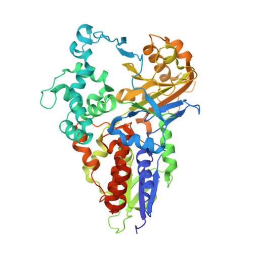
7C3L

Structure of L-lysine oxidase D212A/D315A in complex with L-tyrosine


Experimental Data Snapshot

  • Method: X-RAY DIFFRACTION
  • Resolution: 1.80 Å
  • R-Value Free: 0.190 
  • R-Value Work: 0.159 
  • R-Value Observed: 0.161 

Starting Model: experimental
View more details

wwPDB Validation   3D Report Full Report


Ligand Structure Quality Assessment 


This is version 1.2 of the entry. See complete history


Literature

Structural basis of strict substrate recognition of l-lysine alpha-oxidase from Trichoderma viride.

Kondo, H.Kitagawa, M.Matsumoto, Y.Saito, M.Amano, M.Sugiyama, S.Tamura, T.Kusakabe, H.Inagaki, K.Imada, K.

(2020) Protein Sci 29: 2213-2225

  • DOI: https://doi.org/10.1002/pro.3946
  • Primary Citation of Related Structures:  
    7C3H, 7C3I, 7C3J, 7C3L

  • PubMed Abstract: 

    l-Lysine oxidase (LysOX) is a FAD-dependent homodimeric enzyme that catalyzes the oxidative deamination of l-lysine to produce α-keto-ε-aminocaproate with ammonia and hydrogen peroxide. LysOX shows strict substrate specificity for l-lysine, whereas most l-amino acid oxidases (LAAOs) exhibit broad substrate specificity for l-amino acids. Previous studies of LysOX showed that overall structural similarity to the well-studied snake venom LAAOs. However, the molecular mechanism of strict specificity for l-lysine was still unclear. We here determined the structure of LysOX in complex with l-lysine at 1.7 Å resolution. The structure revealed that the hydrogen bonding network formed by D212, D315, and A440 with two water molecules is responsible for the recognition of the side chain amino group. In addition, a narrow hole formed by five hydrophobic residues in the active site contributes to strict substrate specificity. Mutation studies demonstrated that D212 and D315 are essential for l-lysine recognition, and the D212A/D315A double mutant LysOX showed different substrate specificity from LysOX. Moreover, the structural basis of the substrate specificity change has also been revealed by the structural analysis of the mutant variant and its substrate complexes. These results clearly explain the molecular mechanism of the strict specificity of LysOX and suggest that LysOX is a potential candidate for a template to design LAAOs specific to other l-amino acids.


  • Organizational Affiliation

    Department of Macromolecular Science, Graduate School of Science, Osaka University, Osaka, Japan.


Macromolecules
Find similar proteins by:  (by identity cutoff)  |  3D Structure
Entity ID: 1
MoleculeChains Sequence LengthOrganismDetailsImage
L-lysine oxidase
A, B
540Trichoderma virideMutation(s): 2 
EC: 1.4.3.14
UniProt
Find proteins for A0A0G4DCU0 (Hypocrea rufa)
Explore A0A0G4DCU0 
Go to UniProtKB:  A0A0G4DCU0
Entity Groups  
Sequence Clusters30% Identity50% Identity70% Identity90% Identity95% Identity100% Identity
UniProt GroupA0A0G4DCU0
Sequence Annotations
Expand
  • Reference Sequence
Small Molecules
Ligands 4 Unique
IDChains Name / Formula / InChI Key2D Diagram3D Interactions
FAD
Query on FAD

Download Ideal Coordinates CCD File 
C [auth A],
H [auth B]
FLAVIN-ADENINE DINUCLEOTIDE
C27 H33 N9 O15 P2
VWWQXMAJTJZDQX-UYBVJOGSSA-N
TYR (Subject of Investigation/LOI)
Query on TYR

Download Ideal Coordinates CCD File 
D [auth A],
I [auth B]
TYROSINE
C9 H11 N O3
OUYCCCASQSFEME-QMMMGPOBSA-N
SO4
Query on SO4

Download Ideal Coordinates CCD File 
E [auth A],
J [auth B]
SULFATE ION
O4 S
QAOWNCQODCNURD-UHFFFAOYSA-L
GOL
Query on GOL

Download Ideal Coordinates CCD File 
F [auth A],
G [auth A],
K [auth B],
L [auth B]
GLYCEROL
C3 H8 O3
PEDCQBHIVMGVHV-UHFFFAOYSA-N
Experimental Data & Validation

Experimental Data

  • Method: X-RAY DIFFRACTION
  • Resolution: 1.80 Å
  • R-Value Free: 0.190 
  • R-Value Work: 0.159 
  • R-Value Observed: 0.161 
  • Space Group: C 2 2 21
Unit Cell:
Length ( Å )Angle ( ˚ )
a = 117.09α = 90
b = 170.48β = 90
c = 120.44γ = 90
Software Package:
Software NamePurpose
BSSdata collection
PHENIXrefinement
MOSFLMdata reduction
Aimlessdata scaling
PHASERphasing

Structure Validation

View Full Validation Report


Ligand Structure Quality Assessment 


Entry History & Funding Information

Deposition Data


Funding OrganizationLocationGrant Number
Japan Society for the Promotion of Science (JSPS)Japan24560962

Revision History  (Full details and data files)

  • Version 1.0: 2020-09-23
    Type: Initial release
  • Version 1.1: 2020-11-04
    Changes: Database references
  • Version 1.2: 2023-11-29
    Changes: Data collection, Database references, Refinement description