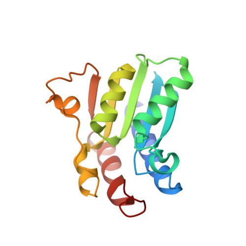
7DCW

The structure of the Arabidopsis thaliana guanosine deaminase complexed with adenosine


Experimental Data Snapshot

  • Method: X-RAY DIFFRACTION
  • Resolution: 2.30 Å
  • R-Value Free: 0.221 
  • R-Value Work: 0.177 
  • R-Value Observed: 0.180 

Starting Model: experimental
View more details

wwPDB Validation   3D Report Full Report


Ligand Structure Quality Assessment 


This is version 1.2 of the entry. See complete history


Literature

Substrate Specificity of GSDA Revealed by Cocrystal Structures and Binding Studies.

Jia, Q.Zhang, J.Zeng, H.Tang, J.Xiao, N.Gao, S.Li, H.Xie, W.

(2022) Int J Mol Sci 23

  • DOI: https://doi.org/10.3390/ijms232314976
  • Primary Citation of Related Structures:  
    7DCB, 7DCW, 7DGC, 7DH1, 7DM5, 7DM6, 7DQN, 7W1Q

  • PubMed Abstract: 

    In plants, guanosine deaminase (GSDA) catalyzes the deamination of guanosine for nitrogen recycling and re-utilization. We previously solved crystal structures of GSDA from Arabidopsis thaliana (AtGSDA) and identified several novel substrates for this enzyme, but the structural basis of the enzyme activation/inhibition is poorly understood. Here, we continued to solve 8 medium-to-high resolution (1.85-2.60 Å) cocrystal structures, which involved AtGSDA and its variants bound by a few ligands, and investigated their binding modes through structural studies and thermal shift analysis. Besides the lack of a 2-amino group of these guanosine derivatives, we discovered that AtGSDA's inactivity was due to the its inability to seclude its active site. Furthermore, the C-termini of the enzyme displayed conformational diversities under certain circumstances. The lack of functional amino groups or poor interactions/geometries of the ligands at the active sites to meet the precise binding and activation requirements for deamination both contributed to AtGSDA's inactivity toward the ligands. Altogether, our combined structural and biochemical studies provide insight into GSDA.


  • Organizational Affiliation

    MOE Key Laboratory of Gene Function and Regulation, School of Life Sciences, Sun Yat-Sen University, Guangzhou 510006, China.


Macromolecules
Find similar proteins by:  (by identity cutoff)  |  3D Structure
Entity ID: 1
MoleculeChains Sequence LengthOrganismDetailsImage
Guanosine deaminaseA,
B [auth D]
161Arabidopsis thalianaMutation(s): 0 
Gene Names: GSDATAD4At5g28050
EC: 3.5.4.15
UniProt
Find proteins for Q94BU8 (Arabidopsis thaliana)
Explore Q94BU8 
Go to UniProtKB:  Q94BU8
Entity Groups  
Sequence Clusters30% Identity50% Identity70% Identity90% Identity95% Identity100% Identity
UniProt GroupQ94BU8
Sequence Annotations
Expand
  • Reference Sequence
Experimental Data & Validation

Experimental Data

  • Method: X-RAY DIFFRACTION
  • Resolution: 2.30 Å
  • R-Value Free: 0.221 
  • R-Value Work: 0.177 
  • R-Value Observed: 0.180 
  • Space Group: P 61
Unit Cell:
Length ( Å )Angle ( ˚ )
a = 119.084α = 90
b = 119.084β = 90
c = 39.773γ = 120
Software Package:
Software NamePurpose
PHENIXrefinement
CrysalisProdata reduction
CrysalisProdata scaling
PHASERphasing

Structure Validation

View Full Validation Report


Ligand Structure Quality Assessment 


Entry History & Funding Information

Deposition Data


Funding OrganizationLocationGrant Number
National Natural Science Foundation of China (NSFC)China31870782

Revision History  (Full details and data files)

  • Version 1.0: 2021-10-27
    Type: Initial release
  • Version 1.1: 2023-02-15
    Changes: Database references
  • Version 1.2: 2023-11-29
    Changes: Data collection, Refinement description