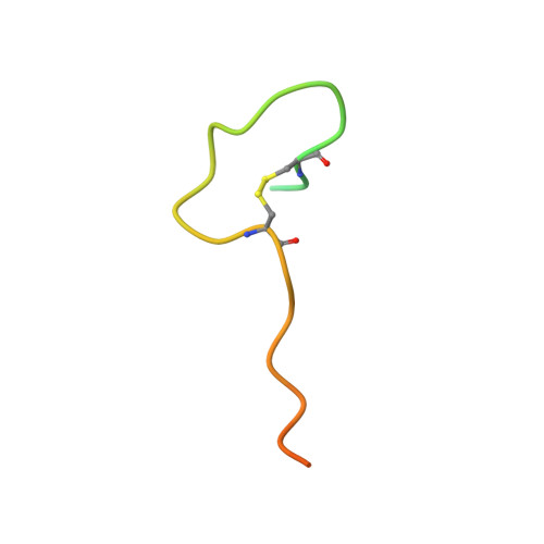
7E6J

Aspartyl/Asparaginyl beta-hydroxylase (AspH) H725A in complex with Factor X peptide fragment (39mer-4Ser)


Experimental Data Snapshot

  • Method: X-RAY DIFFRACTION
  • Resolution: 1.90 Å
  • R-Value Free: 0.213 
  • R-Value Work: 0.170 
  • R-Value Observed: 0.172 

Starting Model: experimental
View more details

wwPDB Validation   3D Report Full Report


Ligand Structure Quality Assessment 


This is version 1.2 of the entry. See complete history


Literature

Human Oxygenase Variants Employing a Single Protein Fe II Ligand Are Catalytically Active.

Brasnett, A.Pfeffer, I.Brewitz, L.Chowdhury, R.Nakashima, Y.Tumber, A.McDonough, M.A.Schofield, C.J.

(2021) Angew Chem Int Ed Engl 60: 14657-14663

  • DOI: https://doi.org/10.1002/anie.202103711
  • Primary Citation of Related Structures:  
    7E6J

  • PubMed Abstract: 

    Aspartate/asparagine-β-hydroxylase (AspH) is a human 2-oxoglutarate (2OG) and Fe II oxygenase that catalyses C3 hydroxylations of aspartate/asparagine residues of epidermal growth factor-like domains (EGFDs). Unusually, AspH employs two histidine residues to chelate Fe II rather than the typical triad of two histidine and one glutamate/aspartate residue. We report kinetic, inhibition, and crystallographic studies concerning human AspH variants in which either of its Fe II binding histidine residues are substituted for alanine. Both the H725A and, in particular, the H679A AspH variants retain substantial catalytic activity. Crystal structures clearly reveal metal-ligation by only a single protein histidine ligand. The results have implications for the functional assignment of 2OG oxygenases and for the design of non-protein biomimetic catalysts.


  • Organizational Affiliation

    Chemistry Research Laboratory and the Ineos Oxford Institute for Antimicrobial Research, University of Oxford, 12 Mansfield Road, Oxford, OX1 3TA, UK.


Macromolecules
Find similar proteins by:  (by identity cutoff)  |  3D Structure
Entity ID: 1
MoleculeChains Sequence LengthOrganismDetailsImage
Aspartyl/asparaginyl beta-hydroxylase429Homo sapiensMutation(s): 1 
Gene Names: ASPHBAH
EC: 1.14.11.16
UniProt & NIH Common Fund Data Resources
Find proteins for Q12797 (Homo sapiens)
Explore Q12797 
Go to UniProtKB:  Q12797
PHAROS:  Q12797
GTEx:  ENSG00000198363 
Entity Groups  
Sequence Clusters30% Identity50% Identity70% Identity90% Identity95% Identity100% Identity
UniProt GroupQ12797
Sequence Annotations
Expand
  • Reference Sequence
Find similar proteins by:  (by identity cutoff)  |  3D Structure
Entity ID: 2
MoleculeChains Sequence LengthOrganismDetailsImage
Peptide from Factor X light chain39Homo sapiensMutation(s): 4 
EC: 3.4.21.6
UniProt & NIH Common Fund Data Resources
Find proteins for P00742 (Homo sapiens)
Explore P00742 
Go to UniProtKB:  P00742
PHAROS:  P00742
GTEx:  ENSG00000126218 
Entity Groups  
Sequence Clusters30% Identity50% Identity70% Identity90% Identity95% Identity100% Identity
UniProt GroupP00742
Sequence Annotations
Expand
  • Reference Sequence
Experimental Data & Validation

Experimental Data

  • Method: X-RAY DIFFRACTION
  • Resolution: 1.90 Å
  • R-Value Free: 0.213 
  • R-Value Work: 0.170 
  • R-Value Observed: 0.172 
  • Space Group: P 21 21 21
Unit Cell:
Length ( Å )Angle ( ˚ )
a = 50.237α = 90
b = 86.841β = 90
c = 123.542γ = 90
Software Package:
Software NamePurpose
PHENIXrefinement
xia2data reduction
xia2data scaling
PHASERphasing

Structure Validation

View Full Validation Report


Ligand Structure Quality Assessment 


Entry History 

Deposition Data

Revision History  (Full details and data files)

  • Version 1.0: 2021-06-30
    Type: Initial release
  • Version 1.1: 2023-11-29
    Changes: Data collection, Database references, Refinement description
  • Version 1.2: 2024-11-20
    Changes: Structure summary