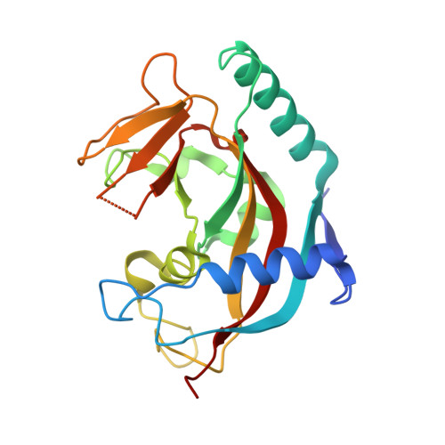
7KKP

Structure of the catalytic domain of tankyrase 1 in complex with niraparib


Experimental Data Snapshot

  • Method: X-RAY DIFFRACTION
  • Resolution: 1.40 Å
  • R-Value Free: 0.208 
  • R-Value Work: 0.187 
  • R-Value Observed: 0.188 

Starting Model: experimental
View more details

wwPDB Validation   3D Report Full Report


Ligand Structure Quality Assessment 


This is version 1.3 of the entry. See complete history


Literature

Dissecting the molecular determinants of clinical PARP1 inhibitor selectivity for tankyrase1.

Ryan, K.Bolanos, B.Smith, M.Palde, P.B.Cuenca, P.D.VanArsdale, T.L.Niessen, S.Zhang, L.Behenna, D.Ornelas, M.A.Tran, K.T.Kaiser, S.Lum, L.Stewart, A.Gajiwala, K.S.

(2021) J Biol Chem 296: 100251-100251

  • DOI: https://doi.org/10.1074/jbc.RA120.016573
  • Primary Citation of Related Structures:  
    7KK2, 7KK3, 7KK4, 7KK5, 7KK6, 7KKM, 7KKN, 7KKO, 7KKP, 7KKQ

  • PubMed Abstract: 

    Poly-ADP-ribosyltransferases play a critical role in DNA repair and cell death, and poly(ADP-ribosyl) polymerase 1 (PARP1) is a particularly important therapeutic target for the treatment of breast cancer because of its synthetic lethal relationship with breast cancer susceptibility proteins 1 and 2. Numerous PARP1 inhibitors have been developed, and their efficacy in cancer treatment is attributed to both the inhibition of enzymatic activity and their ability to trap PARP1 on to the damaged DNA, which is cytotoxic. Of the clinical PARP inhibitors, talazoparib is the most effective at trapping PARP1 on damaged DNA. Biochemically, talazoparib is also suspected to be a potent inhibitor of PARP5a/b (tankyrase1/2 [TNKS1/2]), which is an important regulator of Wnt/β-catenin pathway. Here we show using competition experiments in cell lysate that, at a clinically relevant concentration, talazoparib can potentially bind and engage TNKS1. Using surface plasmon resonance, we measured the dissociation constants of talazoparib, olaparib, niraparib, and veliparib for their interaction with PARP1 and TNKS1. The results show that talazoparib has strong affinity for PARP1 as well as uniquely strong affinity for TNKS1. Finally, we used crystallography and hydrogen deuterium exchange mass spectroscopy to dissect the molecular mechanism of differential selectivity of these PARP1 inhibitors. From these data, we conclude that subtle differences between the ligand-binding sites of PARP1 and TNKS1, differences in the electrostatic nature of the ligands, protein dynamics, and ligand conformational energetics contribute to the different pharmacology of these PARP1 inhibitors. These results will help in the design of drugs to treat Wnt/β-catenin pathway-related cancers, such as colorectal cancers.


  • Organizational Affiliation

    Structural Biology and Protein Science, Pfizer Worldwide Research and Development, San Diego, California, USA.


Macromolecules
Find similar proteins by:  (by identity cutoff)  |  3D Structure
Entity ID: 1
MoleculeChains Sequence LengthOrganismDetailsImage
Poly [ADP-ribose] polymerase
A, B
213Homo sapiensMutation(s): 0 
EC: 2.4.2 (PDB Primary Data), 2.4.2.30 (UniProt)
UniProt & NIH Common Fund Data Resources
Find proteins for O95271 (Homo sapiens)
Explore O95271 
Go to UniProtKB:  O95271
PHAROS:  O95271
GTEx:  ENSG00000173273 
Entity Groups  
Sequence Clusters30% Identity50% Identity70% Identity90% Identity95% Identity100% Identity
UniProt GroupO95271
Sequence Annotations
Expand
  • Reference Sequence
Small Molecules
Ligands 3 Unique
IDChains Name / Formula / InChI Key2D Diagram3D Interactions
3JD (Subject of Investigation/LOI)
Query on 3JD

Download Ideal Coordinates CCD File 
D [auth A],
I [auth B]
2-{4-[(3S)-piperidin-3-yl]phenyl}-2H-indazole-7-carboxamide
C19 H20 N4 O
PCHKPVIQAHNQLW-CQSZACIVSA-N
SO4
Query on SO4

Download Ideal Coordinates CCD File 
E [auth A]
F [auth A]
G [auth A]
J [auth B]
K [auth B]
E [auth A],
F [auth A],
G [auth A],
J [auth B],
K [auth B],
L [auth B],
M [auth B]
SULFATE ION
O4 S
QAOWNCQODCNURD-UHFFFAOYSA-L
ZN
Query on ZN

Download Ideal Coordinates CCD File 
C [auth A],
H [auth B]
ZINC ION
Zn
PTFCDOFLOPIGGS-UHFFFAOYSA-N
Experimental Data & Validation

Experimental Data

  • Method: X-RAY DIFFRACTION
  • Resolution: 1.40 Å
  • R-Value Free: 0.208 
  • R-Value Work: 0.187 
  • R-Value Observed: 0.188 
  • Space Group: P 21 21 21
Unit Cell:
Length ( Å )Angle ( ˚ )
a = 43.37α = 90
b = 74.08β = 90
c = 151.76γ = 90
Software Package:
Software NamePurpose
BUSTERrefinement
PDB_EXTRACTdata extraction
Aimlessdata scaling
PHASERphasing
autoPROCdata reduction

Structure Validation

View Full Validation Report


Ligand Structure Quality Assessment 


Entry History 

Deposition Data

Revision History  (Full details and data files)

  • Version 1.0: 2021-01-06
    Type: Initial release
  • Version 1.1: 2021-01-13
    Changes: Database references
  • Version 1.2: 2021-04-28
    Changes: Database references
  • Version 1.3: 2023-10-18
    Changes: Data collection, Database references, Refinement description