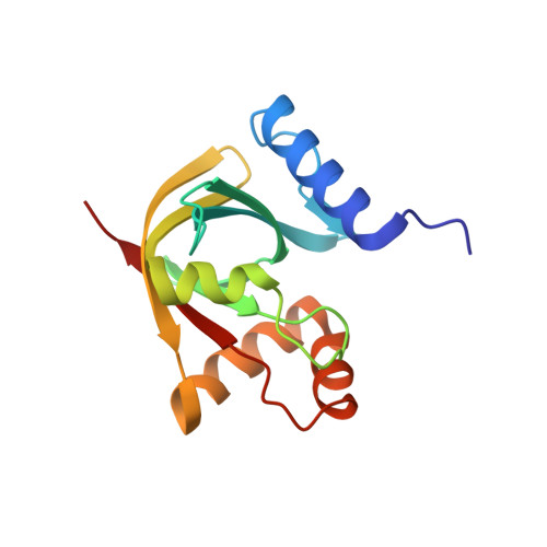
7KL8

Structure of F420 binding protein Rv1558 from Mycobacterium tuberculosis with F420 bound


Experimental Data Snapshot

  • Method: X-RAY DIFFRACTION
  • Resolution: 2.47 Å
  • R-Value Free: 0.220 
  • R-Value Work: 0.200 
  • R-Value Observed: 0.201 

Starting Model: experimental
View more details

wwPDB Validation   3D Report Full Report


Ligand Structure Quality Assessment 


This is version 1.2 of the entry. See complete history


Literature

Potency boost of a Mycobacterium tuberculosis dihydrofolate reductase inhibitor by multienzyme F 420 H 2 -dependent reduction.

Aragaw, W.W.Lee, B.M.Yang, X.Zimmerman, M.D.Gengenbacher, M.Dartois, V.Chui, W.K.Jackson, C.J.Dick, T.

(2021) Proc Natl Acad Sci U S A 118

  • DOI: https://doi.org/10.1073/pnas.2025172118
  • Primary Citation of Related Structures:  
    7KL8

  • PubMed Abstract: 

    Triaza-coumarin (TA-C) is a Mycobacterium tuberculosis (Mtb) dihydrofolate reductase (DHFR) inhibitor with an IC 50 (half maximal inhibitory concentration) of ∼1 µM against the enzyme. Despite this moderate target inhibition, TA-C shows exquisite antimycobacterial activity (MIC 50 , concentration inhibiting growth by 50% = 10 to 20 nM). Here, we investigated the mechanism underlying this potency disconnect. To confirm that TA-C targets DHFR and investigate its unusual potency pattern, we focused on resistance mechanisms. In Mtb, resistance to DHFR inhibitors is frequently associated with mutations in thymidylate synthase thyA , which sensitizes Mtb to DHFR inhibition, rather than in DHFR itself. We observed thyA mutations, consistent with TA-C interfering with the folate pathway. A second resistance mechanism involved biosynthesis of the redox coenzyme F 420 Thus, we hypothesized that TA-C may be metabolized by Mtb F 420 -dependent oxidoreductases (FDORs). By chemically blocking the putative site of FDOR-mediated reduction in TA-C, we reproduced the F 420 -dependent resistance phenotype, suggesting that F 420 H 2 -dependent reduction is required for TA-C to exert its potent antibacterial activity. Indeed, chemically synthesized TA-C-Acid, the putative product of TA-C reduction, displayed a 100-fold lower IC 50 against DHFR. Screening seven recombinant Mtb FDORs revealed that at least two of these enzymes reduce TA-C. This redundancy in activation explains why no mutations in the activating enzymes were identified in the resistance screen. Analysis of the reaction products confirmed that FDORs reduce TA-C at the predicted site, yielding TA-C-Acid. This work demonstrates that intrabacterial metabolism converts TA-C, a moderately active "prodrug," into a 100-fold-more-potent DHFR inhibitor, thus explaining the disconnect between enzymatic and whole-cell activity.


  • Organizational Affiliation

    Center for Discovery and Innovation, Hackensack Meridian Health, Nutley, NJ 07110.


Macromolecules
Find similar proteins by:  (by identity cutoff)  |  3D Structure
Entity ID: 1
MoleculeChains Sequence LengthOrganismDetailsImage
Deazaflavin-dependent nitroreductase
A, B
140Mycobacterium tuberculosisMutation(s): 0 
Gene Names: 
EC: 1 (PDB Primary Data), 1.1.98 (PDB Primary Data)
UniProt
Find proteins for P9WP11 (Mycobacterium tuberculosis (strain ATCC 25618 / H37Rv))
Explore P9WP11 
Go to UniProtKB:  P9WP11
Entity Groups  
Sequence Clusters30% Identity50% Identity70% Identity90% Identity95% Identity100% Identity
UniProt GroupP9WP11
Sequence Annotations
Expand
  • Reference Sequence
Small Molecules
Ligands 3 Unique
IDChains Name / Formula / InChI Key2D Diagram3D Interactions
6J4
Query on 6J4

Download Ideal Coordinates CCD File 
C [auth A]COENZYME F420-3
C34 H43 N6 O21 P
YHDAXCLOUDHUAA-LROHGRLLSA-N
F42
Query on F42

Download Ideal Coordinates CCD File 
H [auth B]COENZYME F420
C29 H36 N5 O18 P
GEHSZWRGPHDXJO-NALJQGANSA-N
SO4
Query on SO4

Download Ideal Coordinates CCD File 
D [auth A]
E [auth A]
F [auth A]
G [auth A]
I [auth B]
D [auth A],
E [auth A],
F [auth A],
G [auth A],
I [auth B],
J [auth B]
SULFATE ION
O4 S
QAOWNCQODCNURD-UHFFFAOYSA-L
Experimental Data & Validation

Experimental Data

  • Method: X-RAY DIFFRACTION
  • Resolution: 2.47 Å
  • R-Value Free: 0.220 
  • R-Value Work: 0.200 
  • R-Value Observed: 0.201 
  • Space Group: P 43 3 2
Unit Cell:
Length ( Å )Angle ( ˚ )
a = 169.544α = 90
b = 169.544β = 90
c = 169.544γ = 90
Software Package:
Software NamePurpose
REFMACrefinement
XDSdata reduction
Aimlessdata scaling
MOLREPphasing

Structure Validation

View Full Validation Report


Ligand Structure Quality Assessment 


Entry History & Funding Information

Deposition Data


Funding OrganizationLocationGrant Number
National Health and Medical Research Council (NHMRC, Australia)AustraliaAPP1128929

Revision History  (Full details and data files)

  • Version 1.0: 2021-03-24
    Type: Initial release
  • Version 1.1: 2021-10-06
    Changes: Database references
  • Version 1.2: 2023-10-18
    Changes: Data collection, Refinement description