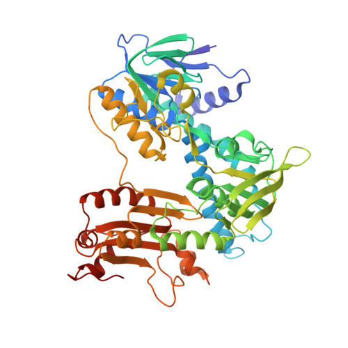
7MGN

Crystal structure of F501H/H506E variant of 2-ketopropyl coenzyme M oxidoreductase/carboxylase (2-KPCC) from Xanthobacter autotrophicus


Experimental Data Snapshot

  • Method: X-RAY DIFFRACTION
  • Resolution: 1.80 Å
  • R-Value Free: 0.189 
  • R-Value Work: 0.141 
  • R-Value Observed: 0.143 

Starting Model: experimental
View more details

wwPDB Validation   3D Report Full Report


Ligand Structure Quality Assessment 


This is version 1.3 of the entry. See complete history


Literature

The unique Phe-His dyad of 2-ketopropyl coenzyme M oxidoreductase/carboxylase selectively promotes carboxylation and S-C bond cleavage.

Prussia, G.A.Shisler, K.A.Zadvornyy, O.A.Streit, B.R.DuBois, J.L.Peters, J.W.

(2021) J Biol Chem 297: 100961-100961

  • DOI: https://doi.org/10.1016/j.jbc.2021.100961
  • Primary Citation of Related Structures:  
    7MGN, 7MGO

  • PubMed Abstract: 

    The 2-ketopropyl-coenzyme M oxidoreductase/carboxylase (2-KPCC) enzyme is the only member of the disulfide oxidoreductase (DSOR) family of enzymes, which are important for reductively cleaving S-S bonds, to have carboxylation activity. 2-KPCC catalyzes the conversion of 2-ketopropyl-coenzyme M to acetoacetate, which is used as a carbon source, in a controlled reaction to exclude protons. A conserved His-Glu motif present in DSORs is key in the protonation step; however, in 2-KPCC, the dyad is substituted by Phe-His. Here, we propose that this difference is important for coupling carboxylation with C-S bond cleavage. We substituted the Phe-His dyad in 2-KPCC to be more DSOR like, replacing the phenylalanine with histidine (F501H) and the histidine with glutamate (H506E), and solved crystal structures of F501H and the double variant F501H_H506E. We found that F501 protects the enolacetone intermediate from protons and that the F501H variant strongly promotes protonation. We also provided evidence for the involvement of the H506 residue in stabilizing the developing charge during the formation of acetoacetate, which acts as a product inhibitor in the WT but not the H506E variant enzymes. Finally, we determined that the F501H substitution promotes a DSOR-like charge transfer interaction with flavin adenine dinucleotide, eliminating the need for cysteine as an internal base. Taken together, these results indicate that the 2-KPCC dyad is responsible for selectively promoting carboxylation and inhibiting protonation in the formation of acetoacetate.


  • Organizational Affiliation

    Institute of Biological Chemistry, Washington State University, Pullman, Washington, USA.


Macromolecules
Find similar proteins by:  (by identity cutoff)  |  3D Structure
Entity ID: 1
MoleculeChains Sequence LengthOrganismDetailsImage
2-oxopropyl-CoM reductase, carboxylating
A, B
523Xanthobacter autotrophicus Py2Mutation(s): 2 
Gene Names: xecCXaut_4867
EC: 1.8.1.5
UniProt
Find proteins for Q56839 (Xanthobacter autotrophicus (strain ATCC BAA-1158 / Py2))
Explore Q56839 
Go to UniProtKB:  Q56839
Entity Groups  
Sequence Clusters30% Identity50% Identity70% Identity90% Identity95% Identity100% Identity
UniProt GroupQ56839
Sequence Annotations
Expand
  • Reference Sequence
Small Molecules
Ligands 3 Unique
IDChains Name / Formula / InChI Key2D Diagram3D Interactions
FDA
Query on FDA

Download Ideal Coordinates CCD File 
D [auth A],
G [auth B]
DIHYDROFLAVINE-ADENINE DINUCLEOTIDE
C27 H35 N9 O15 P2
YPZRHBJKEMOYQH-UYBVJOGSSA-N
COM (Subject of Investigation/LOI)
Query on COM

Download Ideal Coordinates CCD File 
C [auth A],
F [auth B]
1-THIOETHANESULFONIC ACID
C2 H6 O3 S2
ZNEWHQLOPFWXOF-UHFFFAOYSA-N
MG
Query on MG

Download Ideal Coordinates CCD File 
E [auth A],
H [auth B]
MAGNESIUM ION
Mg
JLVVSXFLKOJNIY-UHFFFAOYSA-N
Experimental Data & Validation

Experimental Data

  • Method: X-RAY DIFFRACTION
  • Resolution: 1.80 Å
  • R-Value Free: 0.189 
  • R-Value Work: 0.141 
  • R-Value Observed: 0.143 
  • Space Group: P 1 21 1
Unit Cell:
Length ( Å )Angle ( ˚ )
a = 87.006α = 90
b = 60.111β = 100.52
c = 105.14γ = 90
Software Package:
Software NamePurpose
PHENIXrefinement
XDSdata reduction
XDSdata scaling
PHENIXphasing

Structure Validation

View Full Validation Report


Ligand Structure Quality Assessment 


Entry History & Funding Information

Deposition Data


Funding OrganizationLocationGrant Number
Department of Energy (DOE, United States)United StatesDE-FG0204ER15563

Revision History  (Full details and data files)

  • Version 1.0: 2021-07-21
    Type: Initial release
  • Version 1.1: 2021-07-28
    Changes: Database references
  • Version 1.2: 2021-08-18
    Changes: Advisory, Database references
  • Version 1.3: 2023-10-18
    Changes: Data collection, Refinement description