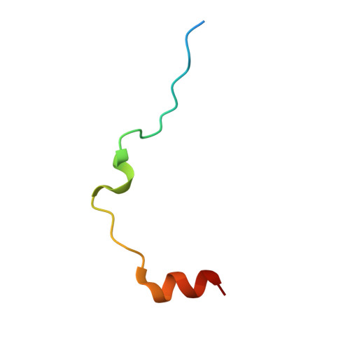
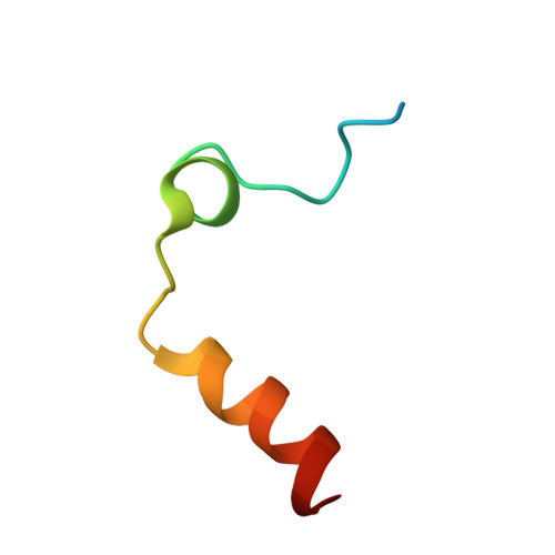
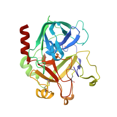
7MJ5

complex of human thrombin with XC-43


Experimental Data Snapshot

  • Method: X-RAY DIFFRACTION
  • Resolution: 2.15 Å
  • R-Value Free: 0.193 
  • R-Value Work: 0.165 
  • R-Value Observed: 0.166 

Starting Model: experimental
View more details

wwPDB Validation   3D Report Full Report


Ligand Structure Quality Assessment 


This is version 1.2 of the entry. See complete history


Literature

Identification of a substrate-like cleavage-resistant thrombin inhibitor from the saliva of the flea Xenopsylla cheopis.

Lu, S.Tirloni, L.Oliveira, M.B.Bosio, C.F.Nardone, G.A.Zhang, Y.Hinnebusch, B.J.Ribeiro, J.M.Andersen, J.F.

(2021) J Biol Chem 297: 101322-101322

  • DOI: https://doi.org/10.1016/j.jbc.2021.101322
  • Primary Citation of Related Structures:  
    7MJ5

  • PubMed Abstract: 

    The salivary glands of the flea Xenopsylla cheopis, a vector of the plague bacterium, Yersinia pestis, express proteins and peptides thought to target the hemostatic and inflammatory systems of its mammalian hosts. Past transcriptomic analyses of salivary gland tissue revealed the presence of two similar peptides (XC-42 and XC-43) having no extensive similarities to any other deposited sequences. Here we show that these peptides specifically inhibit coagulation of plasma and the amidolytic activity of α-thrombin. XC-43, the smaller of the two peptides, is a fast, tight-binding inhibitor of thrombin with a dissociation constant of less than 10 pM. XC-42 exhibits similar selectivity as well as kinetic and binding properties. The crystal structure of XC-43 in complex with thrombin shows that despite its substrate-like binding mode, XC-43 is not detectably cleaved by thrombin and that it interacts with the thrombin surface from the enzyme catalytic site through the fibrinogen-binding exosite I. The low rate of hydrolysis was verified in solution experiments with XC-43, which show the substrate to be largely intact after 2 h of incubation with thrombin at 37 °C. The low rate of XC-43 cleavage by thrombin may be attributable to specific changes in the catalytic triad observable in the crystal structure of the complex or to extensive interactions in the prime sites that may stabilize the binding of cleavage products. Based on the increased arterial occlusion time, tail bleeding time, and blood coagulation parameters in rat models of thrombosis XC-43 could be valuable as an anticoagulant.


  • Organizational Affiliation

    Laboratory of Malaria and Vector Research, National Institute of Allergy and Infectious Diseases, Bethesda, Maryland, USA.


Macromolecules
Find similar proteins by:  (by identity cutoff)  |  3D Structure
Entity ID: 1
MoleculeChains Sequence LengthOrganismDetailsImage
Putative secreted salivary protein
A, N, O, P, Q
A, N, O, P, Q, R
36Xenopsylla cheopisMutation(s): 0 
UniProt
Find proteins for A2IAB2 (Xenopsylla cheopis)
Explore A2IAB2 
Go to UniProtKB:  A2IAB2
Entity Groups  
Sequence Clusters30% Identity50% Identity70% Identity90% Identity95% Identity100% Identity
UniProt GroupA2IAB2
Sequence Annotations
Expand
  • Reference Sequence
Find similar proteins by:  (by identity cutoff)  |  3D Structure
Entity ID: 2
MoleculeChains Sequence LengthOrganismDetailsImage
Thrombin light chain36Homo sapiensMutation(s): 0 
EC: 3.4.21.5
UniProt & NIH Common Fund Data Resources
Find proteins for P00734 (Homo sapiens)
Explore P00734 
Go to UniProtKB:  P00734
PHAROS:  P00734
GTEx:  ENSG00000180210 
Entity Groups  
Sequence Clusters30% Identity50% Identity70% Identity90% Identity95% Identity100% Identity
UniProt GroupP00734
Sequence Annotations
Expand
  • Reference Sequence
Find similar proteins by:  (by identity cutoff)  |  3D Structure
Entity ID: 3
MoleculeChains Sequence LengthOrganismDetailsImage
Thrombin heavy chain259Homo sapiensMutation(s): 0 
EC: 3.4.21.5
UniProt & NIH Common Fund Data Resources
Find proteins for P00734 (Homo sapiens)
Explore P00734 
Go to UniProtKB:  P00734
PHAROS:  P00734
GTEx:  ENSG00000180210 
Entity Groups  
Sequence Clusters30% Identity50% Identity70% Identity90% Identity95% Identity100% Identity
UniProt GroupP00734
Glycosylation
Glycosylation Sites: 1Go to GlyGen: P00734-1
Sequence Annotations
Expand
  • Reference Sequence
Small Molecules
Ligands 2 Unique
IDChains Name / Formula / InChI Key2D Diagram3D Interactions
NAG
Query on NAG

Download Ideal Coordinates CCD File 
AA [auth J]
CA [auth M]
S [auth H]
U [auth C]
W [auth E]
AA [auth J],
CA [auth M],
S [auth H],
U [auth C],
W [auth E],
Y [auth G]
2-acetamido-2-deoxy-beta-D-glucopyranose
C8 H15 N O6
OVRNDRQMDRJTHS-FMDGEEDCSA-N
NA
Query on NA

Download Ideal Coordinates CCD File 
BA [auth J]
DA [auth M]
T [auth H]
V [auth C]
X [auth E]
BA [auth J],
DA [auth M],
T [auth H],
V [auth C],
X [auth E],
Z [auth G]
SODIUM ION
Na
FKNQFGJONOIPTF-UHFFFAOYSA-N
Experimental Data & Validation

Experimental Data

  • Method: X-RAY DIFFRACTION
  • Resolution: 2.15 Å
  • R-Value Free: 0.193 
  • R-Value Work: 0.165 
  • R-Value Observed: 0.166 
  • Space Group: P 21 21 21
Unit Cell:
Length ( Å )Angle ( ˚ )
a = 113.35α = 90
b = 136.32β = 90
c = 193.95γ = 90
Software Package:
Software NamePurpose
PHENIXrefinement
XDSdata reduction
XSCALEdata scaling
PHASERphasing
PDB_EXTRACTdata extraction

Structure Validation

View Full Validation Report


Ligand Structure Quality Assessment 


Entry History 

Deposition Data

Revision History  (Full details and data files)

  • Version 1.0: 2021-12-22
    Type: Initial release
  • Version 1.1: 2023-10-18
    Changes: Data collection, Refinement description
  • Version 1.2: 2024-10-30
    Changes: Structure summary