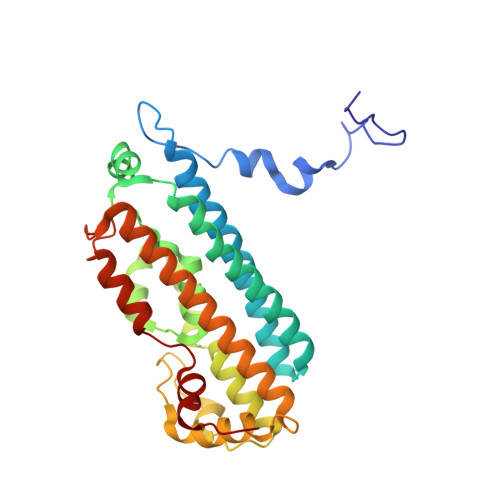
7P46

Crystal Structure of Xanthomonas campestris Tryptophan 2,3-dioxygenase (TDO)


Experimental Data Snapshot

  • Method: X-RAY DIFFRACTION
  • Resolution: 1.70 Å
  • R-Value Free: 0.185 
  • R-Value Work: 0.149 
  • R-Value Observed: 0.151 

Starting Model: experimental
View more details

wwPDB Validation   3D Report Full Report


Ligand Structure Quality Assessment 


This is version 1.1 of the entry. See complete history


Literature

Binding of l-kynurenine to X. campestris tryptophan 2,3-dioxygenase.

Basran, J.Booth, E.S.Campbell, L.P.Thackray, S.J.Jesani, M.H.Clayden, J.Moody, P.C.E.Mowat, C.G.Kwon, H.Raven, E.L.

(2021) J Inorg Biochem 225: 111604-111604

  • DOI: https://doi.org/10.1016/j.jinorgbio.2021.111604
  • Primary Citation of Related Structures:  
    7P46

  • PubMed Abstract: 

    The kynurenine pathway is the major route of tryptophan metabolism. The first step of this pathway is catalysed by one of two heme-dependent dioxygenase enzymes - tryptophan 2,3-dioxygenase (TDO) and indoleamine 2,3-dioxygenase (IDO) - leading initially to the formation of N-formylkynurenine (NFK). In this paper, we present a crystal structure of a bacterial TDO from X. campestris in complex with l-kynurenine, the hydrolysed product of NFK. l-kynurenine is bound at the active site in a similar location to the substrate (l-Trp). Hydrogen bonding interactions with Arg117 and the heme 7-propionate anchor the l-kynurenine molecule into the pocket. A mechanism for the hydrolysis of NFK in the active site is presented.


  • Organizational Affiliation

    Department of Molecular and Cell Biology, Leicester Institute of Structural and Chemical Biology, University of Leicester, Leicester LE1 9HN, UK.


Macromolecules
Find similar proteins by:  (by identity cutoff)  |  3D Structure
Entity ID: 1
MoleculeChains Sequence LengthOrganismDetailsImage
Tryptophan 2,3-dioxygenase
A, B, C, D, E
A, B, C, D, E, F, G, H
282Xanthomonas campestris pv. campestris str. ATCC 33913Mutation(s): 0 
Gene Names: kynAXCC0432
EC: 1.13.11.11
UniProt
Find proteins for Q8PDA8 (Xanthomonas campestris pv. campestris (strain ATCC 33913 / DSM 3586 / NCPPB 528 / LMG 568 / P 25))
Explore Q8PDA8 
Go to UniProtKB:  Q8PDA8
Entity Groups  
Sequence Clusters30% Identity50% Identity70% Identity90% Identity95% Identity100% Identity
UniProt GroupQ8PDA8
Sequence Annotations
Expand
  • Reference Sequence
Small Molecules
Ligands 5 Unique
IDChains Name / Formula / InChI Key2D Diagram3D Interactions
HEM (Subject of Investigation/LOI)
Query on HEM

Download Ideal Coordinates CCD File 
EA [auth D]
I [auth A]
IA [auth E]
LA [auth F]
QA [auth G]
EA [auth D],
I [auth A],
IA [auth E],
LA [auth F],
QA [auth G],
S [auth B],
TA [auth H],
Y [auth C]
PROTOPORPHYRIN IX CONTAINING FE
C34 H32 Fe N4 O4
KABFMIBPWCXCRK-RGGAHWMASA-L
KYN
Query on KYN

Download Ideal Coordinates CCD File 
AA [auth C]
GA [auth D]
JA [auth E]
K [auth A]
NA [auth F]
AA [auth C],
GA [auth D],
JA [auth E],
K [auth A],
NA [auth F],
RA [auth G],
U [auth B],
VA [auth H]
(2S)-2-amino-4-(2-aminophenyl)-4-oxobutanoic acid
C10 H12 N2 O3
YGPSJZOEDVAXAB-QMMMGPOBSA-N
TRP
Query on TRP

Download Ideal Coordinates CCD File 
BA [auth C]
HA [auth D]
KA [auth E]
L [auth A]
OA [auth F]
BA [auth C],
HA [auth D],
KA [auth E],
L [auth A],
OA [auth F],
SA [auth G],
V [auth B],
WA [auth H]
TRYPTOPHAN
C11 H12 N2 O2
QIVBCDIJIAJPQS-VIFPVBQESA-N
GOL
Query on GOL

Download Ideal Coordinates CCD File 
CA [auth C]
DA [auth C]
M [auth A]
N [auth A]
O [auth A]
CA [auth C],
DA [auth C],
M [auth A],
N [auth A],
O [auth A],
P [auth A],
PA [auth F],
Q [auth A],
R [auth A],
W [auth B],
X [auth B],
XA [auth H],
YA [auth H],
ZA [auth H]
GLYCEROL
C3 H8 O3
PEDCQBHIVMGVHV-UHFFFAOYSA-N
CYN
Query on CYN

Download Ideal Coordinates CCD File 
FA [auth D]
J [auth A]
MA [auth F]
T [auth B]
UA [auth H]
FA [auth D],
J [auth A],
MA [auth F],
T [auth B],
UA [auth H],
Z [auth C]
CYANIDE ION
C N
XFXPMWWXUTWYJX-UHFFFAOYSA-N
Experimental Data & Validation

Experimental Data

  • Method: X-RAY DIFFRACTION
  • Resolution: 1.70 Å
  • R-Value Free: 0.185 
  • R-Value Work: 0.149 
  • R-Value Observed: 0.151 
  • Space Group: P 1 21 1
Unit Cell:
Length ( Å )Angle ( ˚ )
a = 78.095α = 90
b = 117.752β = 95.52
c = 138.819γ = 90
Software Package:
Software NamePurpose
REFMACrefinement
PDB_EXTRACTdata extraction
XDSdata reduction
Aimlessdata scaling
PHASERphasing

Structure Validation

View Full Validation Report


Ligand Structure Quality Assessment 


Entry History 

Deposition Data

Revision History  (Full details and data files)

  • Version 1.0: 2021-10-06
    Type: Initial release
  • Version 1.1: 2024-01-31
    Changes: Data collection, Refinement description