7S10

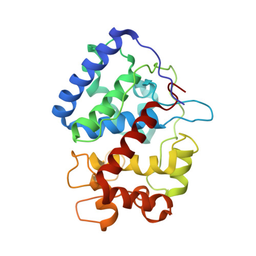
Crystal Structure of ascorbate peroxidase triple mutant: S160M, L203M, Q204M


Experimental Data Snapshot

  • Method: X-RAY DIFFRACTION
  • Resolution: 1.40 Å
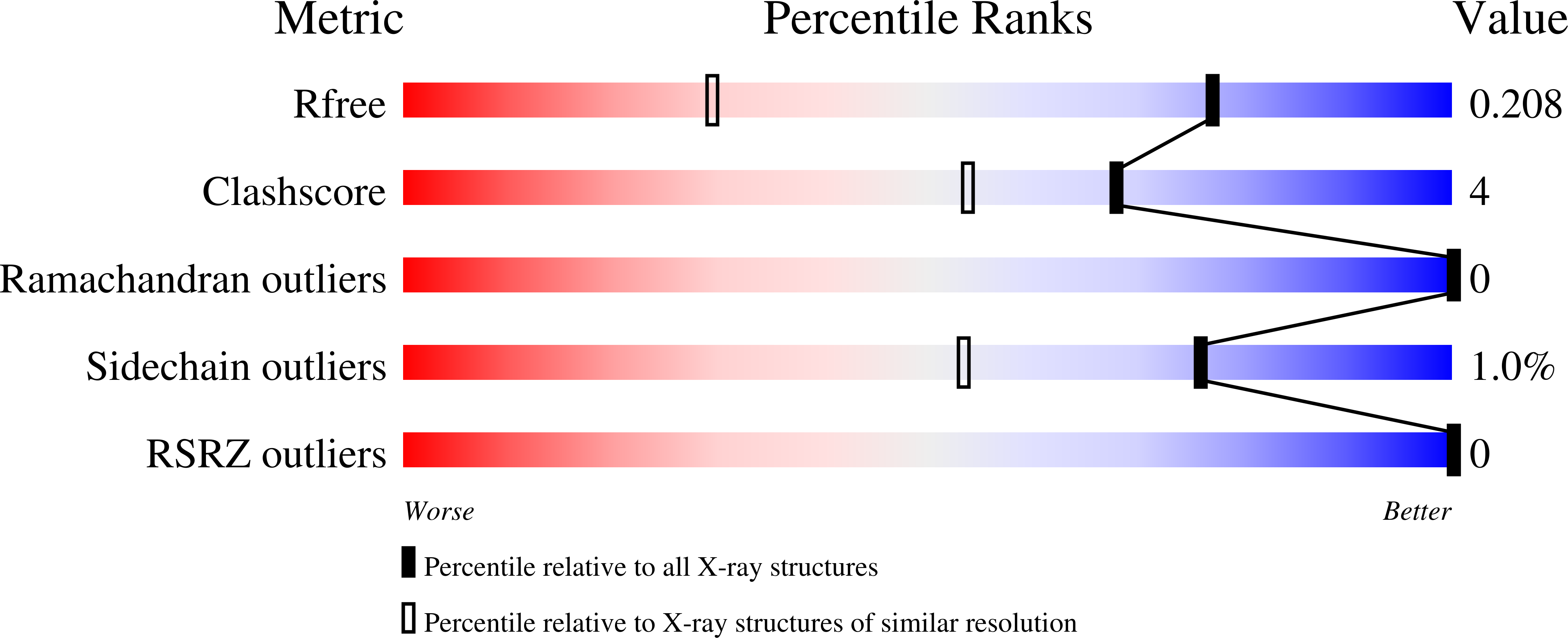
  • R-Value Free: 0.208 
  • R-Value Work: 0.171 
  • R-Value Observed: 0.173 

Starting Model: experimental
View more details

wwPDB Validation   3D Report Full Report


Ligand Structure Quality Assessment 


This is version 1.2 of the entry. See complete history


Literature

Computational analysis of the tryptophan cation radical energetics in peroxidase Compound I.

Poulos, T.L.Kim, J.S.Murarka, V.C.

(2022) J Biol Inorg Chem 27: 229-237

  • DOI: https://doi.org/10.1007/s00775-022-01925-8
  • Primary Citation of Related Structures:  
    7S10

  • PubMed Abstract: 

    Three well-characterized heme peroxidases (cytochrome c peroxidase = CCP, ascorbate peroxidase = APX, and Leishmania major peroxidase = LMP) all have a Trp residue tucked under the heme stacked against the proximal His heme ligand. The reaction of peroxidases with H 2 O 2 to give Compound I results in the oxidation of this Trp to a cationic radical in CCP and LMP but not in APX. Considerable experimental data indicate that the local electrostatic environment controls whether this Trp or the porphyrin is oxidized in Compound I. Attempts have been made to place the differences between these peroxidases on a quantitative basis using computational methods. These efforts have been somewhat limited by the approximations required owing to the computational cost of using fully solvated atomistic models with well-developed forcefields. This now has changed with available GPU computing power and the associated development of software. Here we employ thermodynamic integration and multistate Bennett acceptance ratio methods to help fine-tune our understanding on the energetic differences in Trp radical stabilization in all three peroxidases. These results indicate that the local solvent structure near the redox active Trp plays a significant role in stabilization of the cationic Trp radical.


  • Organizational Affiliation

    Departments of Molecular Biology and Biochemistry, Chemistry, and Pharmaceutical Sciences, University of Calif, Irvine, Irvine, CA, 92697-3900, USA. [email protected].


Macromolecules
Find similar proteins by:  (by identity cutoff)  |  3D Structure
Entity ID: 1
MoleculeChains Sequence LengthOrganismDetailsImage
L-ascorbate peroxidase248Glycine maxMutation(s): 3 
Gene Names: apx1
EC: 1.11.1.11
UniProt
Find proteins for Q43758 (Glycine max)
Explore Q43758 
Go to UniProtKB:  Q43758
Entity Groups  
Sequence Clusters30% Identity50% Identity70% Identity90% Identity95% Identity100% Identity
UniProt GroupQ43758
Sequence Annotations
Expand
  • Reference Sequence
Experimental Data & Validation

Experimental Data

  • Method: X-RAY DIFFRACTION
  • Resolution: 1.40 Å
  • R-Value Free: 0.208 
  • R-Value Work: 0.171 
  • R-Value Observed: 0.173 
  • Space Group: P 42 21 2
Unit Cell:
Length ( Å )Angle ( ˚ )
a = 82.644α = 90
b = 82.644β = 90
c = 75.322γ = 90
Software Package:
Software NamePurpose
PHENIXrefinement
XDSdata reduction
Aimlessdata scaling
PHASERphasing
PDB_EXTRACTdata extraction

Structure Validation

View Full Validation Report


Ligand Structure Quality Assessment 


Entry History & Funding Information

Deposition Data


Funding OrganizationLocationGrant Number
National Institutes of Health/National Institute of General Medical Sciences (NIH/NIGMS)United StatesGM131920

Revision History  (Full details and data files)

  • Version 1.0: 2022-09-07
    Type: Initial release
  • Version 1.1: 2023-10-18
    Changes: Data collection, Refinement description
  • Version 1.2: 2024-09-11
    Changes: Database references, Structure summary