7UY0

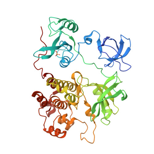
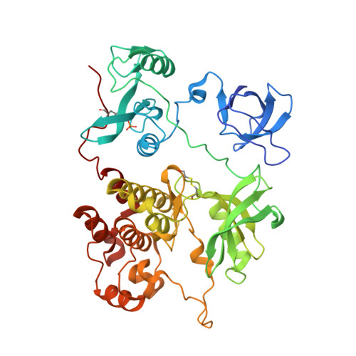
Crystal structure of human Fgr tyrosine kinase in complex with A-419259


Experimental Data Snapshot

  • Method: X-RAY DIFFRACTION
  • Resolution: 2.55 Å
  • R-Value Free: 0.223 
  • R-Value Work: 0.186 
  • R-Value Observed: 0.188 

Starting Model: experimental
View more details

wwPDB Validation   3D Report Full Report


Ligand Structure Quality Assessment 


This is version 1.3 of the entry. See complete history


Literature

ATP-site inhibitors induce unique conformations of the acute myeloid leukemia-associated Src-family kinase, Fgr.

Du, S.Alvarado, J.J.Wales, T.E.Moroco, J.A.Engen, J.R.Smithgall, T.E.

(2022) Structure 30: 1508-1517.e3

  • DOI: https://doi.org/10.1016/j.str.2022.08.008
  • Primary Citation of Related Structures:  
    7UY0, 7UY3

  • PubMed Abstract: 

    The Src-family kinase Fgr is expressed primarily in myeloid hematopoietic cells and contributes to myeloid leukemia. Here, we present X-ray crystal structures of Fgr bound to the ATP-site inhibitors A-419259 and TL02-59, which show promise as anti-leukemic agents. A-419259 induces a closed Fgr conformation, with the SH3 and SH2 domains engaging the SH2-kinase linker and C-terminal tail, respectively. In the Fgr:A-419259 complex, the activation loop of one monomer inserts into the active site of the other, providing a snapshot of trans-autophosphorylation. By contrast, TL02-59 binding induced SH2 domain displacement from the C-terminal tail and SH3 domain release from the linker. Solution studies using HDX MS were consistent with the crystal structures, with A-419259 reducing and TL02-59 enhancing solvent exposure of the SH3 domain. These structures demonstrate that allosteric connections between the kinase and regulatory domains of Src-family kinases are regulated by the ligand bound to the active site.


  • Organizational Affiliation

    Department of Microbiology and Molecular Genetics, University of Pittsburgh School of Medicine, Bridgeside Point II, Suite 523, 450 Technology Drive, Pittsburgh, PA 15219, USA.


Macromolecules
Find similar proteins by:  (by identity cutoff)  |  3D Structure
Entity ID: 1
MoleculeChains Sequence LengthOrganismDetailsImage
Tyrosine-protein kinase Fgr456Homo sapiensMutation(s): 4 
Gene Names: FGRSRC2
EC: 2.7.10.2
UniProt & NIH Common Fund Data Resources
Find proteins for P09769 (Homo sapiens)
Explore P09769 
Go to UniProtKB:  P09769
PHAROS:  P09769
GTEx:  ENSG00000000938 
Entity Groups  
Sequence Clusters30% Identity50% Identity70% Identity90% Identity95% Identity100% Identity
UniProt GroupP09769
Sequence Annotations
Expand
  • Reference Sequence
Find similar proteins by:  (by identity cutoff)  |  3D Structure
Entity ID: 2
MoleculeChains Sequence LengthOrganismDetailsImage
Tyrosine-protein kinase Fgr456Homo sapiensMutation(s): 4 
Gene Names: FGRSRC2
EC: 2.7.10.2
UniProt & NIH Common Fund Data Resources
Find proteins for P09769 (Homo sapiens)
Explore P09769 
Go to UniProtKB:  P09769
PHAROS:  P09769
GTEx:  ENSG00000000938 
Entity Groups  
Sequence Clusters30% Identity50% Identity70% Identity90% Identity95% Identity100% Identity
UniProt GroupP09769
Sequence Annotations
Expand
  • Reference Sequence
Small Molecules
Ligands 4 Unique
IDChains Name / Formula / InChI Key2D Diagram3D Interactions
VSE (Subject of Investigation/LOI)
Query on VSE

Download Ideal Coordinates CCD File 
E [auth A],
H [auth B]
7-[trans-4-(4-methylpiperazin-1-yl)cyclohexyl]-5-(4-phenoxyphenyl)-7H-pyrrolo[2,3-d]pyrimidin-4-amine
C29 H34 N6 O
FDVSOQRNTAPCHB-YHBQERECSA-N
PO4
Query on PO4

Download Ideal Coordinates CCD File 
L [auth B],
M [auth B]
PHOSPHATE ION
O4 P
NBIIXXVUZAFLBC-UHFFFAOYSA-K
GOL
Query on GOL

Download Ideal Coordinates CCD File 
C [auth A],
D [auth A],
G [auth B]
GLYCEROL
C3 H8 O3
PEDCQBHIVMGVHV-UHFFFAOYSA-N
CL
Query on CL

Download Ideal Coordinates CCD File 
F [auth A],
I [auth B],
J [auth B],
K [auth B]
CHLORIDE ION
Cl
VEXZGXHMUGYJMC-UHFFFAOYSA-M
Modified Residues  2 Unique
IDChains TypeFormula2D DiagramParent
PTR
Query on PTR
A
L-PEPTIDE LINKINGC9 H12 N O6 PTYR
CME
Query on CME
B
L-PEPTIDE LINKINGC5 H11 N O3 S2CYS
Experimental Data & Validation

Experimental Data

  • Method: X-RAY DIFFRACTION
  • Resolution: 2.55 Å
  • R-Value Free: 0.223 
  • R-Value Work: 0.186 
  • R-Value Observed: 0.188 
  • Space Group: P 21 21 21
Unit Cell:
Length ( Å )Angle ( ˚ )
a = 49.43α = 90
b = 112.09β = 90
c = 213.73γ = 90
Software Package:
Software NamePurpose
PHENIXrefinement
XDSdata reduction
XSCALEdata scaling
PDB_EXTRACTdata extraction
PHASERphasing

Structure Validation

View Full Validation Report


Ligand Structure Quality Assessment 


Entry History & Funding Information

Deposition Data


Funding OrganizationLocationGrant Number
National Institutes of Health/National Cancer Institute (NIH/NCI)United StatesCA233576

Revision History  (Full details and data files)

  • Version 1.0: 2022-12-28
    Type: Initial release
  • Version 1.1: 2023-10-25
    Changes: Data collection, Refinement description
  • Version 1.2: 2023-11-15
    Changes: Data collection
  • Version 1.3: 2024-11-13
    Changes: Structure summary