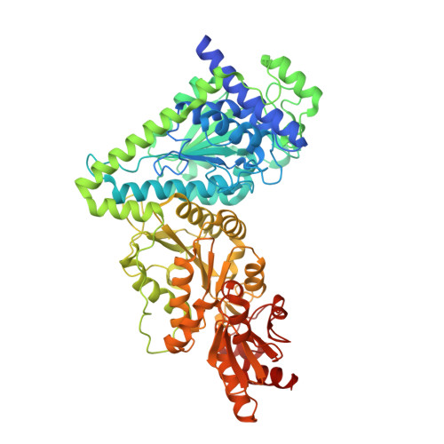
7WRT

X-ray structure ofThermus thermophilus HB8 transketorase demonstrate in complex with TPP and D-erythrose-4-phosphate


Experimental Data Snapshot

  • Method: X-RAY DIFFRACTION
  • Resolution: 2.25 Å
  • R-Value Free: 0.254 
  • R-Value Work: 0.202 

Starting Model: experimental
View more details

wwPDB Validation   3D Report Full Report


Ligand Structure Quality Assessment 


This is version 1.2 of the entry. See complete history


Literature

Structural and biochemical characterizations of Thermus thermophilus HB8 transketolase producing a heptulose.

Yoshihara, A.Takamatsu, Y.Mochizuki, S.Yoshida, H.Masui, R.Izumori, K.Kamitori, S.

(2023) Appl Microbiol Biotechnol 107: 233-245

  • DOI: https://doi.org/10.1007/s00253-022-12297-z
  • Primary Citation of Related Structures:  
    7WRR, 7WRT

  • PubMed Abstract: 

    Transketolase is a key enzyme in the pentose phosphate pathway in all organisms, recognizing sugar phosphates as substrates. Transketolase with a cofactor of thiamine pyrophosphate catalyzes the transfer of a 2-carbon unit from D-xylulose-5-phosphate to D-ribose-5-phosphate (5-carbon aldose), giving D-sedoheptulose-7-phosphate (7-carbon ketose). Transketolases can also recognize non-phosphorylated monosaccharides as substrates, and catalyze the formation of non-phosphorylated 7-carbon ketose (heptulose), which has attracted pharmaceutical attention as an inhibitor of sugar metabolism. Here, we report the structural and biochemical characterizations of transketolase from Thermus thermophilus HB8 (TtTK), a well-characterized thermophilic Gram-negative bacterium. TtTK showed marked thermostability with maximum enzyme activity at 85 °C, and efficiently catalyzed the formation of heptuloses from lithium hydroxypyruvate and four aldopentoses: D-ribose, L-lyxose, L-arabinose, and D-xylose. The X-ray structure showed that TtTK tightly forms a homodimer with more interactions between subunits compared with transketolase from other organisms, contributing to its thermal stability. A modeling study based on X-ray structures suggested that D-ribose and L-lyxose could bind to the catalytic site of TtTK to form favorable hydrogen bonds with the enzyme, explaining the high conversion rates of 41% (D-ribose) and 43% (L-lyxose) to heptulose. These results demonstrate the potential of TtTK as an enzyme producing a rare sugar of heptulose. KEY POINTS: • Transketolase catalyzes the formation of a 7-carbon sugar phosphate • Structural and biochemical characterizations of thermophilic transketolase were done • The enzyme could produce non-phosphorylated 7-carbon ketoses from sugars.


  • Organizational Affiliation

    International Institute of Rare Sugar Research and Education, Kagawa University, Takamatsu, Kagawa, 760-8521, Japan. [email protected].


Macromolecules
Find similar proteins by:  (by identity cutoff)  |  3D Structure
Entity ID: 1
MoleculeChains Sequence LengthOrganismDetailsImage
Transketolase
A, B, C, D
672Thermus thermophilus HB8Mutation(s): 0 
Gene Names: TTHA0108
EC: 2.2.1.1
UniProt
Find proteins for Q5SM35 (Thermus thermophilus (strain ATCC 27634 / DSM 579 / HB8))
Explore Q5SM35 
Go to UniProtKB:  Q5SM35
Entity Groups  
Sequence Clusters30% Identity50% Identity70% Identity90% Identity95% Identity100% Identity
UniProt GroupQ5SM35
Sequence Annotations
Expand
  • Reference Sequence
Small Molecules
Ligands 3 Unique
IDChains Name / Formula / InChI Key2D Diagram3D Interactions
TPP
Query on TPP

Download Ideal Coordinates CCD File 
E [auth A],
I [auth B],
K [auth C],
O [auth D]
THIAMINE DIPHOSPHATE
C12 H19 N4 O7 P2 S
AYEKOFBPNLCAJY-UHFFFAOYSA-O
E4P
Query on E4P

Download Ideal Coordinates CCD File 
G [auth A],
H [auth B],
M [auth C],
N [auth D]
ERYTHOSE-4-PHOSPHATE
C4 H9 O7 P
NGHMDNPXVRFFGS-IUYQGCFVSA-N
CA
Query on CA

Download Ideal Coordinates CCD File 
F [auth A],
J [auth B],
L [auth C],
P [auth D]
CALCIUM ION
Ca
BHPQYMZQTOCNFJ-UHFFFAOYSA-N
Experimental Data & Validation

Experimental Data

  • Method: X-RAY DIFFRACTION
  • Resolution: 2.25 Å
  • R-Value Free: 0.254 
  • R-Value Work: 0.202 
  • Space Group: P 1
Unit Cell:
Length ( Å )Angle ( ˚ )
a = 72.68α = 72.308
b = 88.86β = 88.696
c = 117.37γ = 73.459
Software Package:
Software NamePurpose
REFMACrefinement
XDSdata reduction
XDSdata scaling
MOLREPphasing

Structure Validation

View Full Validation Report


Ligand Structure Quality Assessment 


Entry History & Funding Information

Deposition Data


Funding OrganizationLocationGrant Number
Not funded--

Revision History  (Full details and data files)

  • Version 1.0: 2022-12-07
    Type: Initial release
  • Version 1.1: 2022-12-28
    Changes: Database references
  • Version 1.2: 2023-11-29
    Changes: Data collection, Refinement description