7ZBP

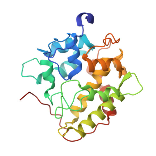
Unspecific peroxygenase from Marasmius rotula


Experimental Data Snapshot

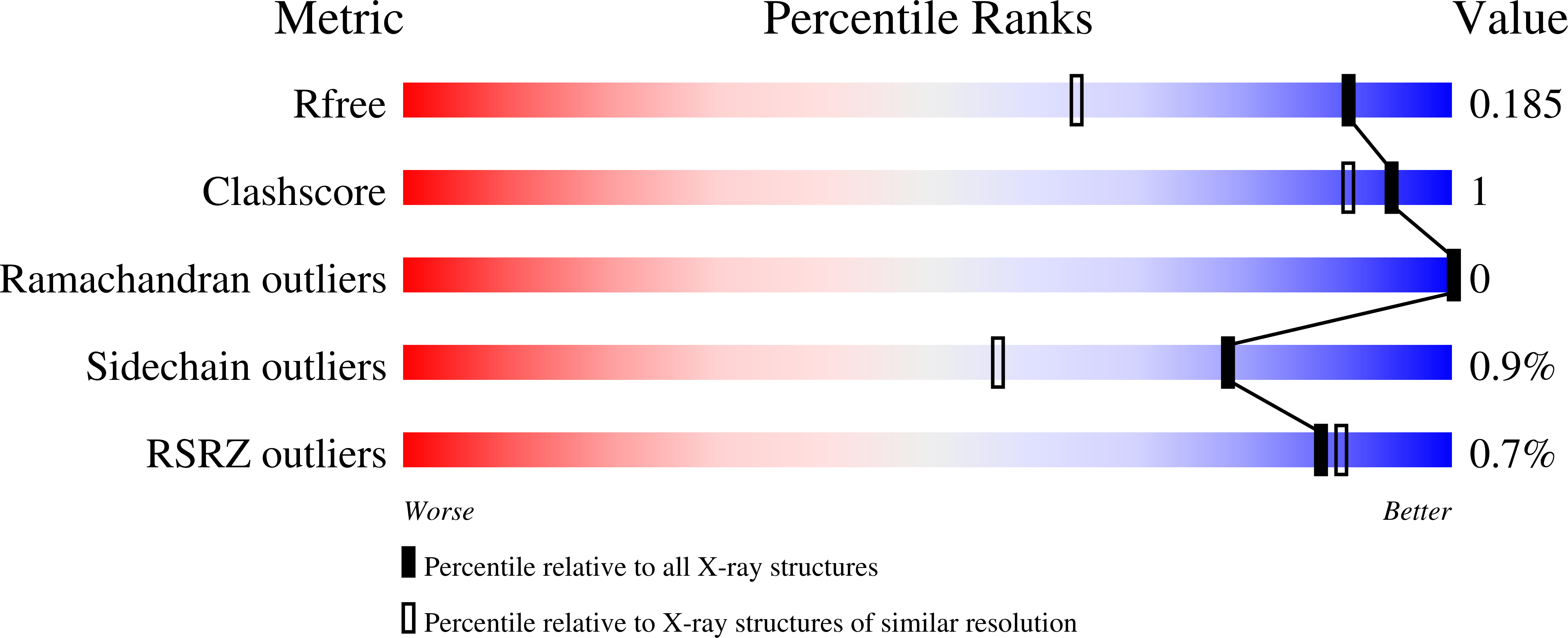
  • Method: X-RAY DIFFRACTION
  • Resolution: 1.45 Å
  • R-Value Free: 0.184 
  • R-Value Work: 0.158 
  • R-Value Observed: 0.159 

Starting Model: experimental
View more details

wwPDB Validation   3D Report Full Report


Ligand Structure Quality Assessment 


This is version 1.3 of the entry. See complete history


Literature

Structural Characterization of Two Short Unspecific Peroxygenases: Two Different Dimeric Arrangements.

Linde, D.Santillana, E.Fernandez-Fueyo, E.Gonzalez-Benjumea, A.Carro, J.Gutierrez, A.Martinez, A.T.Romero, A.

(2022) Antioxidants (Basel) 11

  • DOI: https://doi.org/10.3390/antiox11050891
  • Primary Citation of Related Structures:  
    7ZBP, 7ZCL

  • PubMed Abstract: 

    Unspecific peroxygenases (UPOs) are extracellular fungal enzymes of biotechnological interest as self-sufficient (and more stable) counterparts of cytochrome P450 monooxygenases, the latter being present in most living cells. Expression hosts and structural information are crucial for exploiting UPO diversity (over eight thousand UPO-type genes were identified in sequenced genomes) in target reactions of industrial interest. However, while many thousands of entries in the Protein Data Bank include molecular coordinates of P450 enzymes, only 19 entries correspond to UPO enzymes, and UPO structures from only two species ( Agrocybe aegerita and Hypoxylon sp.) have been published to date. In the present study, two UPOs from the basidiomycete Marasmius rotula (r Mro UPO) and the ascomycete Collariella virescens (r Cvi UPO) were crystallized after sequence optimization and Escherichia coli expression as active soluble enzymes. Crystals of r Mro UPO and r Cvi UPO were obtained at sufficiently high resolution (1.45 and 1.95 Å, respectively) and the corresponding structures were solved by molecular replacement. The crystal structures of the two enzymes (and two mutated variants) showed dimeric proteins. Complementary biophysical and molecular biology studies unveiled the diverse structural bases of the dimeric nature of the two enzymes. Intermolecular disulfide bridge and parallel association between two α-helices, among other interactions, were identified at the dimer interfaces. Interestingly, one of the r Cvi UPO variants incorporated the ability to produce fatty acid diepoxides-reactive compounds with valuable cross-linking capabilities-due to removal of the enzyme C-terminal tail located near the entrance of the heme access channel. In conclusion, different dimeric arrangements could be described in (short) UPO crystal structures.


  • Organizational Affiliation

    Centro de Investigaciones Biológicas "Margarita Salas" (CIB), CSIC, E-28040 Madrid, Spain.


Macromolecules
Find similar proteins by:  (by identity cutoff)  |  3D Structure
Entity ID: 1
MoleculeChains Sequence LengthOrganismDetailsImage
Marasmius rotula UPO
A, B, C, D
239Marasmius rotulaMutation(s): 0 
EC: 1.11.2.1
Entity Groups  
Sequence Clusters30% Identity50% Identity70% Identity90% Identity95% Identity100% Identity
Sequence Annotations
Expand
  • Reference Sequence
Small Molecules
Ligands 5 Unique
IDChains Name / Formula / InChI Key2D Diagram3D Interactions
HEM (Subject of Investigation/LOI)
Query on HEM

Download Ideal Coordinates CCD File 
G [auth A],
J [auth B],
N [auth C],
Q [auth D]
PROTOPORPHYRIN IX CONTAINING FE
C34 H32 Fe N4 O4
KABFMIBPWCXCRK-RGGAHWMASA-L
PEG
Query on PEG

Download Ideal Coordinates CCD File 
E [auth A],
F [auth A]
DI(HYDROXYETHYL)ETHER
C4 H10 O3
MTHSVFCYNBDYFN-UHFFFAOYSA-N
GOL
Query on GOL

Download Ideal Coordinates CCD File 
M [auth B]GLYCEROL
C3 H8 O3
PEDCQBHIVMGVHV-UHFFFAOYSA-N
ACT
Query on ACT

Download Ideal Coordinates CCD File 
I [auth A],
L [auth B],
P [auth C],
S [auth D]
ACETATE ION
C2 H3 O2
QTBSBXVTEAMEQO-UHFFFAOYSA-M
MG
Query on MG

Download Ideal Coordinates CCD File 
H [auth A],
K [auth B],
O [auth C],
R [auth D]
MAGNESIUM ION
Mg
JLVVSXFLKOJNIY-UHFFFAOYSA-N
Experimental Data & Validation

Experimental Data

  • Method: X-RAY DIFFRACTION
  • Resolution: 1.45 Å
  • R-Value Free: 0.184 
  • R-Value Work: 0.158 
  • R-Value Observed: 0.159 
  • Space Group: C 2 2 21
Unit Cell:
Length ( Å )Angle ( ˚ )
a = 100.349α = 90
b = 107.44β = 90
c = 186.231γ = 90
Software Package:
Software NamePurpose
REFMACrefinement
PDB_EXTRACTdata extraction
XDSdata reduction
Aimlessdata scaling
PHASERphasing

Structure Validation

View Full Validation Report


Ligand Structure Quality Assessment 


Entry History & Funding Information

Deposition Data


Funding OrganizationLocationGrant Number
Spanish National Research CouncilSpainPIE-202020E224

Revision History  (Full details and data files)

  • Version 1.0: 2022-05-18
    Type: Initial release
  • Version 1.1: 2022-06-08
    Changes: Database references
  • Version 1.2: 2024-01-31
    Changes: Data collection, Refinement description
  • Version 1.3: 2024-11-06
    Changes: Structure summary