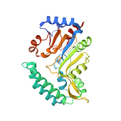
7ZEH

Human cytosolic 5' nucleotidase IIIB in complex with 3,4-diF-Bn7Guanine


Experimental Data Snapshot

  • Method: X-RAY DIFFRACTION
  • Resolution: 1.50 Å
  • R-Value Free: 0.220 
  • R-Value Work: 0.188 
  • R-Value Observed: 0.189 

Starting Model: experimental
View more details

wwPDB Validation   3D Report Full Report


Ligand Structure Quality Assessment 


This is version 1.3 of the entry. See complete history


Literature

Substrate-Based Design of Cytosolic Nucleotidase IIIB Inhibitors and Structural Insights into Inhibition Mechanism.

Kubacka, D.Kozarski, M.Baranowski, M.R.Wojcik, R.Panecka-Hofman, J.Strzelecka, D.Basquin, J.Jemielity, J.Kowalska, J.

(2022) Pharmaceuticals (Basel) 15

  • DOI: https://doi.org/10.3390/ph15050554
  • Primary Citation of Related Structures:  
    7ZEE, 7ZEG, 7ZEH

  • PubMed Abstract: 

    Cytosolic nucleotidases (cNs) catalyze dephosphorylation of nucleoside 5'-monophosphates and thereby contribute to the regulation of nucleotide levels in cells. cNs have also been shown to dephosphorylate several therapeutically relevant nucleotide analogues. cN-IIIB has shown in vitro a distinctive activity towards 7-mehtylguanosine monophosphate (m 7 GMP), which is one key metabolites of mRNA cap. Consequently, it has been proposed that cN-IIIB participates in mRNA cap turnover and prevents undesired accumulation and salvage of m 7 GMP. Here, we sought to develop molecular tools enabling more advanced studies on the cellular role of cN-IIIB. To that end, we performed substrate and inhibitor property profiling using a library of 41 substrate analogs. The most potent hit compounds (identified among m 7 GMP analogs) were used as a starting point for structure-activity relationship studies. As a result, we identified several 7-benzylguanosine 5'-monophosphate (Bn 7 GMP) derivatives as potent, unhydrolyzable cN-IIIB inhibitors. The mechanism of inhibition was elucidated using X-ray crystallography and molecular docking. Finally, we showed that compounds that potently inhibit recombinant cN-IIIB have the ability to inhibit m 7 GMP decay in cell lysates.


  • Organizational Affiliation

    Division of Biophysics, Institute of Experimental Physics, Faculty of Physics, University of Warsaw, Pasteura 5, 02-093 Warsaw, Poland.


Macromolecules
Find similar proteins by:  (by identity cutoff)  |  3D Structure
Entity ID: 1
MoleculeChains Sequence LengthOrganismDetailsImage
7-methylguanosine phosphate-specific 5'-nucleotidase
A, B, C, D
302Homo sapiensMutation(s): 0 
Gene Names: NT5C3BNT5C3L
EC: 3.1.3.91 (PDB Primary Data), 3.1.3.5 (PDB Primary Data)
UniProt & NIH Common Fund Data Resources
Find proteins for Q969T7 (Homo sapiens)
Explore Q969T7 
Go to UniProtKB:  Q969T7
PHAROS:  Q969T7
GTEx:  ENSG00000141698 
Entity Groups  
Sequence Clusters30% Identity50% Identity70% Identity90% Identity95% Identity100% Identity
UniProt GroupQ969T7
Sequence Annotations
Expand
  • Reference Sequence
Small Molecules
Ligands 6 Unique
IDChains Name / Formula / InChI Key2D Diagram3D Interactions
IPQ (Subject of Investigation/LOI)
Query on IPQ

Download Ideal Coordinates CCD File 
E [auth A],
P [auth B],
PA [auth D],
Z [auth C]
N7-(3,4-difluorobenzyl) guanine
C12 H10 F2 N5 O
NBTRICCNNMIXFE-UHFFFAOYSA-O
RBL (Subject of Investigation/LOI)
Query on RBL

Download Ideal Coordinates CCD File 
AA [auth C],
F [auth A],
Q [auth B],
QA [auth D]
D-ribulose
C5 H10 O5
ZAQJHHRNXZUBTE-NQXXGFSBSA-N
SO4
Query on SO4

Download Ideal Coordinates CCD File 
AB [auth D],
NA [auth C],
O [auth A],
Y [auth B]
SULFATE ION
O4 S
QAOWNCQODCNURD-UHFFFAOYSA-L
PO4
Query on PO4

Download Ideal Coordinates CCD File 
MA [auth C],
N [auth A],
X [auth B],
ZA [auth D]
PHOSPHATE ION
O4 P
NBIIXXVUZAFLBC-UHFFFAOYSA-K
EDO
Query on EDO

Download Ideal Coordinates CCD File 
BA [auth C]
CA [auth C]
DA [auth C]
EA [auth C]
FA [auth C]
BA [auth C],
CA [auth C],
DA [auth C],
EA [auth C],
FA [auth C],
G [auth A],
GA [auth C],
H [auth A],
HA [auth C],
I [auth A],
IA [auth C],
J [auth A],
JA [auth C],
K [auth A],
KA [auth C],
L [auth A],
OA [auth D],
R [auth B],
RA [auth D],
S [auth B],
SA [auth D],
T [auth B],
TA [auth D],
U [auth B],
UA [auth D],
V [auth B],
VA [auth D],
WA [auth D],
XA [auth D]
1,2-ETHANEDIOL
C2 H6 O2
LYCAIKOWRPUZTN-UHFFFAOYSA-N
MG
Query on MG

Download Ideal Coordinates CCD File 
LA [auth C],
M [auth A],
W [auth B],
YA [auth D]
MAGNESIUM ION
Mg
JLVVSXFLKOJNIY-UHFFFAOYSA-N
Experimental Data & Validation

Experimental Data

  • Method: X-RAY DIFFRACTION
  • Resolution: 1.50 Å
  • R-Value Free: 0.220 
  • R-Value Work: 0.188 
  • R-Value Observed: 0.189 
  • Space Group: P 1
Unit Cell:
Length ( Å )Angle ( ˚ )
a = 60.59α = 82.219
b = 80.89β = 72.462
c = 83.17γ = 68.202
Software Package:
Software NamePurpose
PHENIXrefinement
XDSdata reduction
XSCALEdata scaling
PHASERphasing

Structure Validation

View Full Validation Report


Ligand Structure Quality Assessment 


Entry History & Funding Information

Deposition Data


Funding OrganizationLocationGrant Number
Polish National Science CentrePolandUMO- 2017/24/C/NZ1/00169

Revision History  (Full details and data files)

  • Version 1.0: 2022-05-25
    Type: Initial release
  • Version 1.1: 2022-06-08
    Changes: Database references
  • Version 1.2: 2024-01-31
    Changes: Data collection, Refinement description
  • Version 1.3: 2024-11-06
    Changes: Structure summary