8A4D

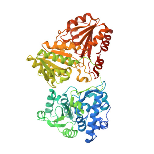
1-deoxy-D-xylulose 5-phosphate synthase from Pseudomonas aeruginosa with a thiamine analog inhibitor


Experimental Data Snapshot

  • Method: X-RAY DIFFRACTION
  • Resolution: 2.20 Å
  • R-Value Free: 0.241 
  • R-Value Work: 0.176 
  • R-Value Observed: 0.179 

Starting Model: experimental
View more details

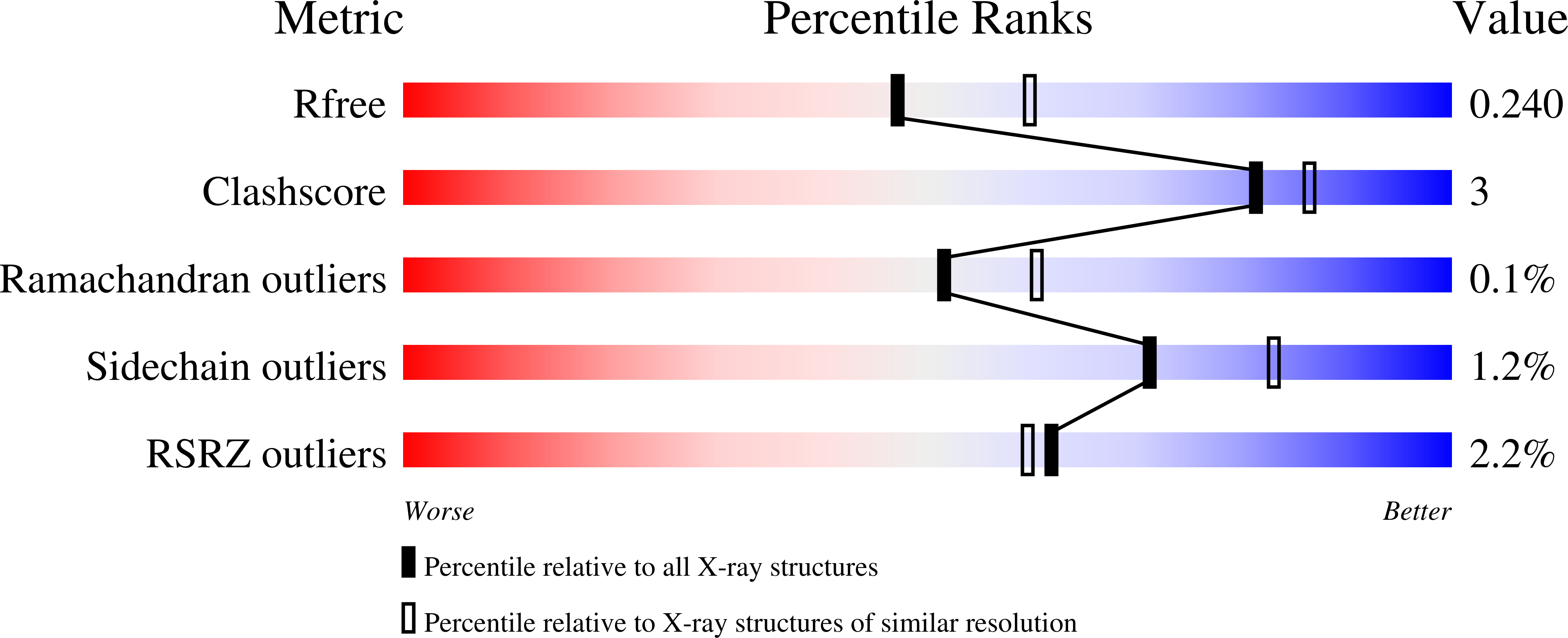
wwPDB Validation   3D Report Full Report


Ligand Structure Quality Assessment 


This is version 1.4 of the entry. See complete history


Literature

1-deoxy-D-xylulose-5-phosphate synthase from Pseudomonas aeruginosa and Klebsiella pneumoniae reveals conformational changes upon cofactor binding.

Hamid, R.Adam, S.Lacour, A.Monjas, L.Kohnke, J.Hirsch, A.K.H.

(2023) J Biol Chem 299: 105152-105152

  • DOI: https://doi.org/10.1016/j.jbc.2023.105152
  • Primary Citation of Related Structures:  
    8A29, 8A45, 8A4D, 8A5K

  • PubMed Abstract: 

    The ESKAPE bacteria are the six highly virulent and antibiotic-resistant pathogens that require the most urgent attention for the development of novel antibiotics. Detailed knowledge of target proteins specific to bacteria is essential to develop novel treatment options. The methylerythritol-phosphate (MEP) pathway, which is absent in humans, represents a potentially valuable target for the development of novel antibiotics. Within the MEP pathway, the enzyme 1-deoxy-D-xylulose-5-phosphate synthase (DXPS) catalyzes a crucial, rate-limiting first step and a branch point in the biosynthesis of the vitamins B1 and B6. We report the high-resolution crystal structures of DXPS from the important ESKAPE pathogens Pseudomonas aeruginosa and Klebsiella pneumoniae in both the co-factor-bound and the apo forms. We demonstrate that the absence of the cofactor thiamine diphosphate results in conformational changes that lead to disordered loops close to the active site that might be important for the design of potent DXPS inhibitors. Collectively, our results provide important structural details that aid in the assessment of DXPS as a potential target in the ongoing efforts to combat antibiotic resistance.


  • Organizational Affiliation

    Department of Drug Design and Optimization, Helmholtz Institute for Pharmaceutical Research Saarland (HIPS) - Helmholtz Centre for Infection Research (HZI), Saarbrücken, Germany; Department of Pharmacy, Saarland University, Saarbrücken, Germany.


Macromolecules
Find similar proteins by:  (by identity cutoff)  |  3D Structure
Entity ID: 1
MoleculeChains Sequence LengthOrganismDetailsImage
1-deoxy-D-xylulose-5-phosphate synthase
A, B, C, D, E
A, B, C, D, E, F
622Pseudomonas aeruginosa LESB58Mutation(s): 0 
Gene Names: dxsPLES_09321
EC: 2.2.1.7
UniProt
Find proteins for B7V7R4 (Pseudomonas aeruginosa (strain LESB58))
Explore B7V7R4 
Go to UniProtKB:  B7V7R4
Entity Groups  
Sequence Clusters30% Identity50% Identity70% Identity90% Identity95% Identity100% Identity
UniProt GroupB7V7R4
Sequence Annotations
Expand
  • Reference Sequence
Small Molecules
Ligands 5 Unique
IDChains Name / Formula / InChI Key2D Diagram3D Interactions
26G (Subject of Investigation/LOI)
Query on 26G

Download Ideal Coordinates CCD File 
G [auth A]
K [auth B]
N [auth C]
S [auth D]
V [auth E]
G [auth A],
K [auth B],
N [auth C],
S [auth D],
V [auth E],
Y [auth F]
2-{3-[(4-amino-2-methylpyrimidin-5-yl)methyl]phenyl}ethanol
C14 H17 N3 O
CLAXVJRRXGSGJP-UHFFFAOYSA-N
GOL
Query on GOL

Download Ideal Coordinates CCD File 
Z [auth F]GLYCEROL
C3 H8 O3
PEDCQBHIVMGVHV-UHFFFAOYSA-N
CA
Query on CA

Download Ideal Coordinates CCD File 
DA [auth F],
M [auth B]
CALCIUM ION
Ca
BHPQYMZQTOCNFJ-UHFFFAOYSA-N
MG
Query on MG

Download Ideal Coordinates CCD File 
AA [auth F]
BA [auth F]
CA [auth F]
H [auth A]
I [auth A]
AA [auth F],
BA [auth F],
CA [auth F],
H [auth A],
I [auth A],
L [auth B],
O [auth C],
P [auth C],
T [auth D],
W [auth E],
X [auth E]
MAGNESIUM ION
Mg
JLVVSXFLKOJNIY-UHFFFAOYSA-N
NA
Query on NA

Download Ideal Coordinates CCD File 
EA [auth F],
J [auth A],
Q [auth C],
R [auth C],
U [auth D]
SODIUM ION
Na
FKNQFGJONOIPTF-UHFFFAOYSA-N
Experimental Data & Validation

Experimental Data

  • Method: X-RAY DIFFRACTION
  • Resolution: 2.20 Å
  • R-Value Free: 0.241 
  • R-Value Work: 0.176 
  • R-Value Observed: 0.179 
  • Space Group: P 21 21 21
Unit Cell:
Length ( Å )Angle ( ˚ )
a = 116.618α = 90
b = 137.618β = 90
c = 231.681γ = 90
Software Package:
Software NamePurpose
PHENIXrefinement
PHENIXrefinement
Aimlessdata scaling
PHASERphasing

Structure Validation

View Full Validation Report


Ligand Structure Quality Assessment 


Entry History & Funding Information

Deposition Data


Funding OrganizationLocationGrant Number
Helmholtz AssociationGermany--

Revision History  (Full details and data files)

  • Version 1.0: 2023-07-12
    Type: Initial release
  • Version 1.1: 2023-08-23
    Changes: Data collection, Database references
  • Version 1.2: 2023-09-06
    Changes: Structure summary
  • Version 1.3: 2023-09-20
    Changes: Database references
  • Version 1.4: 2024-02-07
    Changes: Refinement description