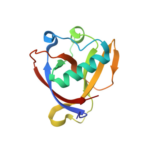
Two Ligand-Binding Sites on SARS-CoV-2 Non-Structural Protein 1 Revealed by Fragment-Based X-ray Screening.
Ma, S., Damfo, S., Lou, J., Pinotsis, N., Bowler, M.W., Haider, S., Kozielski, F.(2022) Int J Mol Sci 23
- PubMed: 36293303
- DOI: https://doi.org/10.3390/ijms232012448
- Primary Citation of Related Structures:
8A55, 8AYS, 8AZ8 - PubMed Abstract:
The regular reappearance of coronavirus (CoV) outbreaks over the past 20 years has caused significant health consequences and financial burdens worldwide. The most recent and still ongoing novel CoV pandemic, caused by Severe Acute Respiratory Syndrome coronavirus 2 (SARS-CoV-2) has brought a range of devastating consequences. Due to the exceptionally fast development of vaccines, the mortality rate of the virus has been curbed to a significant extent. However, the limitations of vaccination efficiency and applicability, coupled with the still high infection rate, emphasise the urgent need for discovering safe and effective antivirals against SARS-CoV-2 by suppressing its replication or attenuating its virulence. Non-structural protein 1 (nsp1), a unique viral and conserved leader protein, is a crucial virulence factor for causing host mRNA degradation, suppressing interferon (IFN) expression and host antiviral signalling pathways. In view of the essential role of nsp1 in the CoV life cycle, it is regarded as an exploitable target for antiviral drug discovery. Here, we report a variety of fragment hits against the N-terminal domain of SARS-CoV-2 nsp1 identified by fragment-based screening via X-ray crystallography. We also determined the structure of nsp1 at atomic resolution (0.99 Å). Binding affinities of hits against nsp1 and potential stabilisation were determined by orthogonal biophysical assays such as microscale thermophoresis and thermal shift assays. We identified two ligand-binding sites on nsp1, one deep and one shallow pocket, which are not conserved between the three medically relevant SARS, SARS-CoV-2 and MERS coronaviruses. Our study provides an excellent starting point for the development of more potent nsp1-targeting inhibitors and functional studies on SARS-CoV-2 nsp1.
Organizational Affiliation:
School of Pharmacy, University College London, 29-39 Brunswick Square, London WC1N 1AX, UK.


