8AOO

Fucosylated mixed-chirality linear peptide FHP31 bound to the fucose binding lectin LecB PA-IIL from Pseudomonas aeruginosa at 1.2 Angstrom resolution.


Experimental Data Snapshot

  • Method: X-RAY DIFFRACTION
  • Resolution: 1.18 Å
  • R-Value Free: 0.151 
  • R-Value Work: 0.127 
  • R-Value Observed: 0.128 

Starting Model: experimental
View more details

wwPDB Validation   3D Report Full Report


Ligand Structure Quality Assessment 


This is version 1.3 of the entry. See complete history


Literature

To Fold or Not to Fold: Diastereomeric Optimization of an alpha-Helical Antimicrobial Peptide.

Personne, H.Paschoud, T.Fulgencio, S.Baeriswyl, S.Kohler, T.van Delden, C.Stocker, A.Javor, S.Reymond, J.L.

(2023) J Med Chem 66: 7570-7583

  • DOI: https://doi.org/10.1021/acs.jmedchem.3c00460
  • Primary Citation of Related Structures:  
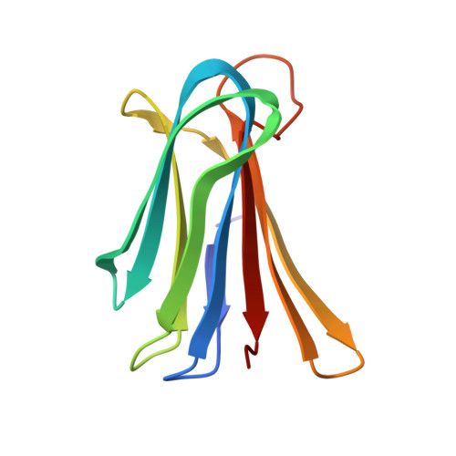
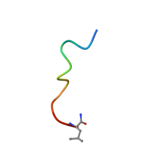
    8AN9, 8ANO, 8ANR, 8AOO

  • PubMed Abstract: 

    Membrane disruptive α-helical antimicrobial peptides (AMPs) offer an opportunity to address multidrug resistance; however, most AMPs are toxic and unstable in serum. These limitations can be partly overcome by introducing D-residues, which often confers protease resistance and reduces toxicity without affecting antibacterial activity, presumably due to lowered α-helicity. Here, we investigated 31 diastereomers of the α-helical AMP KKLLKLLKLLL. Three diastereomers containing two, three, and four D-residues showed increased antibacterial effects, comparable hemolysis, reduced toxicity against HEK293 cells, and excellent serum stability, while another diastereomer with four D-residues additionally displayed lower hemolysis. X-ray crystallography confirmed that high or low α-helicity as measured by circular dichroism indicated α-helical or disordered structures independently of the number of chirality switched residues. In contrast to previous reports, α-helicity across diastereomers correlated with both antibacterial activity and hemolysis and revealed a complex relationship between stereochemistry, activity, and toxicity, highlighting the potential of diastereomers for property optimization.


  • Organizational Affiliation

    Department of Chemistry, Biochemistry and Pharmaceutical Sciences, University of Bern, Freiestrasse 3, CH-3012 Bern, Switzerland.


Macromolecules
Find similar proteins by:  (by identity cutoff)  |  3D Structure
Entity ID: 1
MoleculeChains Sequence LengthOrganismDetailsImage
Fucose-binding lectin PA-IIL
A, B, C, D
114Pseudomonas aeruginosa PAO1Mutation(s): 0 
Gene Names: lecBPA3361
UniProt
Find proteins for Q9HYN5 (Pseudomonas aeruginosa (strain ATCC 15692 / DSM 22644 / CIP 104116 / JCM 14847 / LMG 12228 / 1C / PRS 101 / PAO1))
Explore Q9HYN5 
Go to UniProtKB:  Q9HYN5
Entity Groups  
Sequence Clusters30% Identity50% Identity70% Identity90% Identity95% Identity100% Identity
UniProt GroupQ9HYN5
Sequence Annotations
Expand
  • Reference Sequence

Find similar proteins by:  Sequence   |   3D Structure  

Entity ID: 2
MoleculeChains Sequence LengthOrganismDetailsImage
Fucosylated mixed-chirality peptide FHP31E,
F,
G [auth H],
H [auth I]
12synthetic constructMutation(s): 0 
Sequence Annotations
Expand
  • Reference Sequence
Small Molecules
Ligands 2 Unique
IDChains Name / Formula / InChI Key2D Diagram3D Interactions
ZDC (Subject of Investigation/LOI)
Query on ZDC

Download Ideal Coordinates CCD File 
Q [auth E],
R [auth F],
S [auth H],
T [auth I]
3,7-anhydro-2,8-dideoxy-L-glycero-D-gluco-octonic acid
C8 H14 O6
YTZUDUWVQZSKNN-OASCRQMUSA-N
CA
Query on CA

Download Ideal Coordinates CCD File 
I [auth A]
J [auth A]
K [auth B]
L [auth B]
M [auth C]
I [auth A],
J [auth A],
K [auth B],
L [auth B],
M [auth C],
N [auth C],
O [auth D],
P [auth D]
CALCIUM ION
Ca
BHPQYMZQTOCNFJ-UHFFFAOYSA-N
Experimental Data & Validation

Experimental Data

  • Method: X-RAY DIFFRACTION
  • Resolution: 1.18 Å
  • R-Value Free: 0.151 
  • R-Value Work: 0.127 
  • R-Value Observed: 0.128 
  • Space Group: P 1 21 1
Unit Cell:
Length ( Å )Angle ( ˚ )
a = 52.777α = 90
b = 62.67β = 93.19
c = 64.407γ = 90
Software Package:
Software NamePurpose
XDSdata reduction
XSCALEdata scaling
PHASERphasing
PHENIXrefinement
PDB_EXTRACTdata extraction

Structure Validation

View Full Validation Report


Ligand Structure Quality Assessment 


Entry History & Funding Information

Deposition Data


Funding OrganizationLocationGrant Number
European Research Council (ERC)European Union--
Swiss National Science FoundationSwitzerland--

Revision History  (Full details and data files)

  • Version 1.0: 2023-05-31
    Type: Initial release
  • Version 1.1: 2023-06-07
    Changes: Database references
  • Version 1.2: 2023-06-21
    Changes: Database references
  • Version 1.3: 2024-02-07
    Changes: Data collection, Refinement description