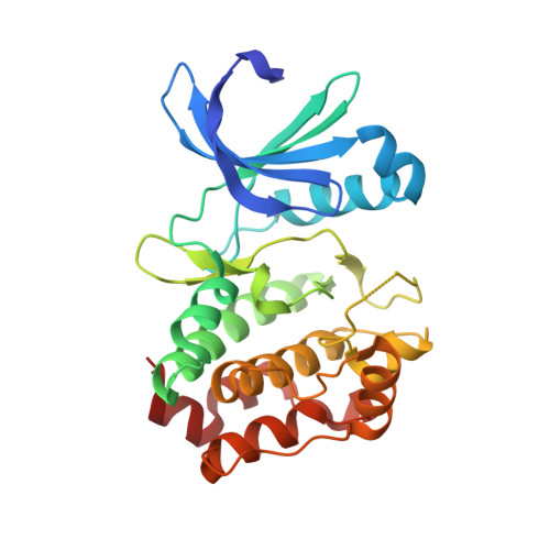
8C1D

Aurora A kinase in complex with TPX2-inhibitor 9


Experimental Data Snapshot

  • Method: X-RAY DIFFRACTION
  • Resolution: 2.12 Å
  • R-Value Free: 0.238 
  • R-Value Work: 0.210 
  • R-Value Observed: 0.212 

Starting Model: experimental
View more details

wwPDB Validation   3D Report Full Report


Ligand Structure Quality Assessment 


This is version 1.2 of the entry. See complete history


Literature

Selective Aurora A-TPX2 Interaction Inhibitors Have In Vivo Efficacy as Targeted Antimitotic Agents.

Stockwell, S.R.Scott, D.E.Fischer, G.Guarino, E.Rooney, T.P.C.Feng, T.S.Moschetti, T.Srinivasan, R.Alza, E.Asteian, A.Dagostin, C.Alcaide, A.Rocaboy, M.Blaszczyk, B.Higueruelo, A.Wang, X.Rossmann, M.Perrior, T.R.Blundell, T.L.Spring, D.R.McKenzie, G.Abell, C.Skidmore, J.Venkitaraman, A.R.Hyvonen, M.

(2024) J Med Chem 67: 15521-15536

  • DOI: https://doi.org/10.1021/acs.jmedchem.4c01165
  • Primary Citation of Related Structures:  
    8C14, 8C15, 8C1D, 8C1E, 8C1F, 8C1G, 8C1H, 8C1I, 8C1K, 8C1M

  • PubMed Abstract: 

    Aurora A kinase, a cell division regulator, is frequently overexpressed in various cancers, provoking genome instability and resistance to antimitotic chemotherapy. Localization and enzymatic activity of Aurora A are regulated by its interaction with the spindle assembly factor TPX2. We have used fragment-based, structure-guided lead discovery to develop small molecule inhibitors of the Aurora A-TPX2 protein-protein interaction (PPI). Our lead compound, CAM2602 , inhibits Aurora A:TPX2 interaction, binding Aurora A with 19 nM affinity. CAM2602 exhibits oral bioavailability, causes pharmacodynamic biomarker modulation, and arrests the growth of tumor xenografts. CAM2602 acts by a novel mechanism compared to ATP-competitive inhibitors and is highly specific to Aurora A over Aurora B. Consistent with our finding that Aurora A overexpression drives taxane resistance, these inhibitors synergize with paclitaxel to suppress the outgrowth of pancreatic cancer cells. Our results provide a blueprint for targeting the Aurora A-TPX2 PPI for cancer therapy and suggest a promising clinical utility for this mode of action.


  • Organizational Affiliation

    Medical Research Council Cancer Unit, University of Cambridge, Cambridge CB2 0XZ, U.K.


Macromolecules
Find similar proteins by:  (by identity cutoff)  |  3D Structure
Entity ID: 1
MoleculeChains Sequence LengthOrganismDetailsImage
Aurora kinase A265Homo sapiensMutation(s): 1 
Gene Names: AURKAAIKAIRK1ARK1AURAAYK1BTAKIAK1STK15STK6
EC: 2.7.11.1
UniProt & NIH Common Fund Data Resources
Find proteins for O14965 (Homo sapiens)
Explore O14965 
Go to UniProtKB:  O14965
PHAROS:  O14965
GTEx:  ENSG00000087586 
Entity Groups  
Sequence Clusters30% Identity50% Identity70% Identity90% Identity95% Identity100% Identity
UniProt GroupO14965
Sequence Annotations
Expand
  • Reference Sequence
Small Molecules
Ligands 7 Unique
IDChains Name / Formula / InChI Key2D Diagram3D Interactions
ADP
Query on ADP

Download Ideal Coordinates CCD File 
C [auth A]ADENOSINE-5'-DIPHOSPHATE
C10 H15 N5 O10 P2
XTWYTFMLZFPYCI-KQYNXXCUSA-N
T2F (Subject of Investigation/LOI)
Query on T2F

Download Ideal Coordinates CCD File 
B [auth A]4-(4-chlorophenyl)-7-methyl-1~{H}-indole-6-carboxylic acid
C16 H12 Cl N O2
HXZMDXUYYVDPRW-UHFFFAOYSA-N
T5L
Query on T5L

Download Ideal Coordinates CCD File 
F [auth A](1~{R},2~{R})-cyclohexane-1,2-dicarboxylic acid
C8 H12 O4
QSAWQNUELGIYBC-PHDIDXHHSA-N
SO4
Query on SO4

Download Ideal Coordinates CCD File 
G [auth A]
H [auth A]
I [auth A]
J [auth A]
K [auth A]
G [auth A],
H [auth A],
I [auth A],
J [auth A],
K [auth A],
L [auth A]
SULFATE ION
O4 S
QAOWNCQODCNURD-UHFFFAOYSA-L
DMS
Query on DMS

Download Ideal Coordinates CCD File 
M [auth A],
N [auth A]
DIMETHYL SULFOXIDE
C2 H6 O S
IAZDPXIOMUYVGZ-UHFFFAOYSA-N
ACT
Query on ACT

Download Ideal Coordinates CCD File 
O [auth A]ACETATE ION
C2 H3 O2
QTBSBXVTEAMEQO-UHFFFAOYSA-M
MG
Query on MG

Download Ideal Coordinates CCD File 
D [auth A],
E [auth A]
MAGNESIUM ION
Mg
JLVVSXFLKOJNIY-UHFFFAOYSA-N
Experimental Data & Validation

Experimental Data

  • Method: X-RAY DIFFRACTION
  • Resolution: 2.12 Å
  • R-Value Free: 0.238 
  • R-Value Work: 0.210 
  • R-Value Observed: 0.212 
  • Space Group: P 61 2 2
Unit Cell:
Length ( Å )Angle ( ˚ )
a = 81.341α = 90
b = 81.341β = 90
c = 171.531γ = 120
Software Package:
Software NamePurpose
Aimlessdata scaling
BUSTERrefinement
PDB_EXTRACTdata extraction
XDSdata reduction
PHASERphasing

Structure Validation

View Full Validation Report


Ligand Structure Quality Assessment 


Entry History & Funding Information

Deposition Data


Funding OrganizationLocationGrant Number
Wellcome TrustUnited Kingdom090340/Z/09/Z "101134/Z/13/Z

Revision History  (Full details and data files)

  • Version 1.0: 2024-01-10
    Type: Initial release
  • Version 1.1: 2024-09-04
    Changes: Database references
  • Version 1.2: 2024-09-25
    Changes: Database references