8ELA

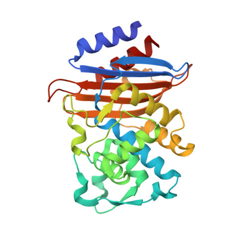
CTX-M-14 beta-lactamase mutant - N132A w MES


Experimental Data Snapshot

  • Method: X-RAY DIFFRACTION
  • Resolution: 1.50 Å
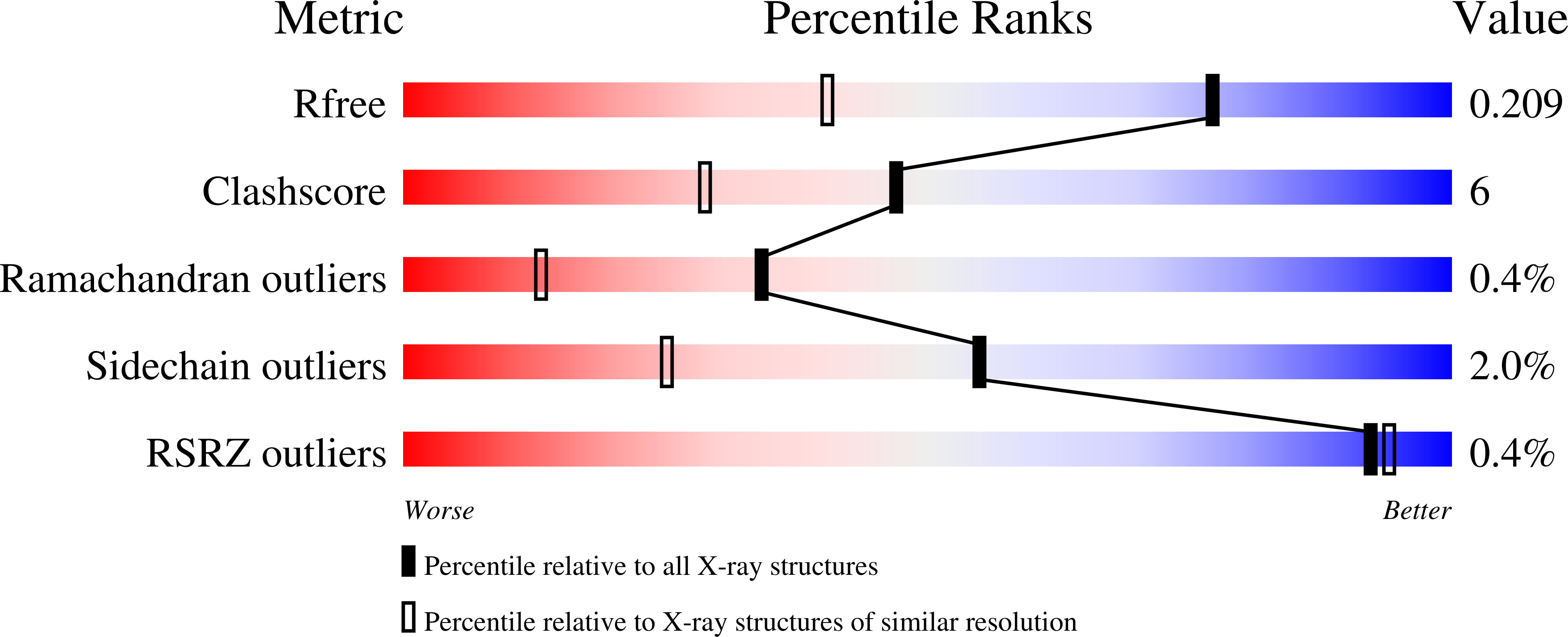
  • R-Value Free: 0.209 
  • R-Value Work: 0.173 
  • R-Value Observed: 0.175 

Starting Model: experimental
View more details

wwPDB Validation   3D Report Full Report


Ligand Structure Quality Assessment 


This is version 1.2 of the entry. See complete history


Literature

Mutagenesis and structural analysis reveal the CTX-M beta-lactamase active site is optimized for cephalosporin catalysis and drug resistance.

Lu, S.Montoya, M.Hu, L.Neetu, N.Sankaran, B.Prasad, B.V.V.Palzkill, T.

(2023) J Biol Chem 299: 104630-104630

  • DOI: https://doi.org/10.1016/j.jbc.2023.104630
  • Primary Citation of Related Structures:  
    8DOD, 8DOE, 8DON, 8DP4, 8DPQ, 8ELA, 8ELB

  • PubMed Abstract: 

    CTX-M β-lactamases are a widespread source of resistance to β-lactam antibiotics in Gram-negative bacteria. These enzymes readily hydrolyze penicillins and cephalosporins, including oxyimino-cephalosporins such as cefotaxime. To investigate the preference of CTX-M enzymes for cephalosporins, we examined eleven active-site residues in the CTX-M-14 β-lactamase model system by alanine mutagenesis to assess the contribution of the residues to catalysis and specificity for the hydrolysis of the penicillin, ampicillin, and the cephalosporins cephalothin and cefotaxime. Key active site residues for class A β-lactamases, including Lys73, Ser130, Asn132, Lys234, Thr216, and Thr235, contribute significantly to substrate binding and catalysis of penicillin and cephalosporin substrates in that alanine substitutions decrease both k cat and k cat /K M . A second group of residues, including Asn104, Tyr105, Asn106, Thr215, and Thr216, contribute only to substrate binding, with the substitutions decreasing only k cat /K M . Importantly, calculating the average effect of a substitution across the 11 active-site residues shows that the most significant impact is on cefotaxime hydrolysis while ampicillin hydrolysis is least affected, suggesting the active site is highly optimized for cefotaxime catalysis. Furthermore, we determined X-ray crystal structures for the apo-enzymes of the mutants N106A, S130A, N132A, N170A, T215A, and T235A. Surprisingly, in the structures of some mutants, particularly N106A and T235A, the changes in structure propagate from the site of substitution to other regions of the active site, suggesting that the impact of substitutions is due to more widespread changes in structure and illustrating the interconnected nature of the active site.


  • Organizational Affiliation

    Department of Pharmacology and Chemical Biology, Baylor College of Medicine, Houston, Texas, USA.


Macromolecules
Find similar proteins by:  (by identity cutoff)  |  3D Structure
Entity ID: 1
MoleculeChains Sequence LengthOrganismDetailsImage
Beta-lactamase
A, B
263Escherichia coliMutation(s): 1 
Gene Names: 
EC: 3.5.2.6
UniProt
Find proteins for Q9L5C7 (Escherichia coli)
Explore Q9L5C7 
Go to UniProtKB:  Q9L5C7
Entity Groups  
Sequence Clusters30% Identity50% Identity70% Identity90% Identity95% Identity100% Identity
UniProt GroupQ9L5C7
Sequence Annotations
Expand
  • Reference Sequence
Small Molecules
Ligands 4 Unique
IDChains Name / Formula / InChI Key2D Diagram3D Interactions
MES (Subject of Investigation/LOI)
Query on MES

Download Ideal Coordinates CCD File 
C [auth A],
G [auth B]
2-(N-MORPHOLINO)-ETHANESULFONIC ACID
C6 H13 N O4 S
SXGZJKUKBWWHRA-UHFFFAOYSA-N
SO4
Query on SO4

Download Ideal Coordinates CCD File 
D [auth A],
H [auth B]
SULFATE ION
O4 S
QAOWNCQODCNURD-UHFFFAOYSA-L
ZN
Query on ZN

Download Ideal Coordinates CCD File 
E [auth A],
I [auth B]
ZINC ION
Zn
PTFCDOFLOPIGGS-UHFFFAOYSA-N
CL
Query on CL

Download Ideal Coordinates CCD File 
F [auth A],
J [auth B]
CHLORIDE ION
Cl
VEXZGXHMUGYJMC-UHFFFAOYSA-M
Experimental Data & Validation

Experimental Data

  • Method: X-RAY DIFFRACTION
  • Resolution: 1.50 Å
  • R-Value Free: 0.209 
  • R-Value Work: 0.173 
  • R-Value Observed: 0.175 
  • Space Group: C 2 2 21
Unit Cell:
Length ( Å )Angle ( ˚ )
a = 59.314α = 90
b = 60.43β = 90
c = 264.216γ = 90
Software Package:
Software NamePurpose
SCALAdata scaling
PHENIXrefinement
PDB_EXTRACTdata extraction
iMOSFLMdata reduction
PHASERphasing

Structure Validation

View Full Validation Report


Ligand Structure Quality Assessment 


Entry History & Funding Information

Deposition Data


Funding OrganizationLocationGrant Number
National Institutes of Health/National Institute Of Allergy and Infectious Diseases (NIH/NIAID)United StatesAI32956

Revision History  (Full details and data files)

  • Version 1.0: 2023-04-05
    Type: Initial release
  • Version 1.1: 2023-04-26
    Changes: Database references
  • Version 1.2: 2023-10-25
    Changes: Data collection, Refinement description