The oxidation of cholesterol derivatives by the CYP124 and CYP142 enzymes from Mycobacterium marinum.
Ghith, A., Bruning, J.B., Bell, S.G.(2023) J Steroid Biochem Mol Biol 231: 106317-106317
- PubMed: 37141947
- DOI: https://doi.org/10.1016/j.jsbmb.2023.106317
- Primary Citation of Related Structures:
8GDI - PubMed Abstract:
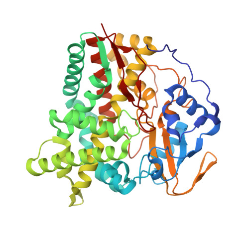
The CYP124 and CYP142 families of bacterial cytochrome P450 monooxygenases (CYPs), catalyze the oxidation of methyl branched lipids, including cholesterol, as one of the initial activating steps in their catabolism. Both enzymes are reported to supplement the CYP125 family of P450 enzymes. These CYP125 enzymes are found in the same bacteria, and are the primary cholesterol/cholest-4-en-3-one metabolizing enzymes. To further understand the role of the CYP124 and CYP142 cytochrome P450s we investigated the Mycobacterium marinum enzymes, MmarCYP124A1 and CYP142A3, with various cholesterol analogs with modifications on the A and B rings of the steroid. We assessed the substrate binding and catalytic activity of each enzyme. Neither enzyme could bind or oxidize cholesteryl acetate or 3,5-cholestadiene, which have modifications at the C3 hydroxyl moiety of cholesterol. The CYP142 enzyme was better able to accommodate and oxidize cholesterol analogs which have changes on the A/B rings including cholesterol-5α,6α-epoxide and diastereomers of 5-cholestan-3-ol. The CYP124 enzyme was more tolerant of changes at C7 of the cholesterol B ring, e.g., 7-ketocholesterol than in the A ring. The selectivity for oxidation at the ω-carbon of a branched chain was observed in all steroids that were oxidized. The 7-ketocholesterol-bound MmarCYP124A1 enzyme from M. marinum, was structurally characterized by X-ray crystallography to 1.81 Å resolution. The 7-ketocholesterol-bound X-ray crystal structure of the MmarCYP124A1 enzyme revealed that the substrate binding mode of this cholesterol derivative was altered compared to those observed with other non-steroidal ligands. The structure provided an explanation for the selectivity of the enzyme for terminal methyl hydroxylation.
Organizational Affiliation:
Department of Chemistry, University of Adelaide, SA 5005, Australia.


