8JQP

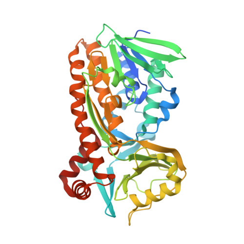
Protocatecuate hydroxylase from Xylophilus ampelinus complexed with 3,4-dihydroxybenzoate


Experimental Data Snapshot

  • Method: X-RAY DIFFRACTION
  • Resolution: 1.65 Å
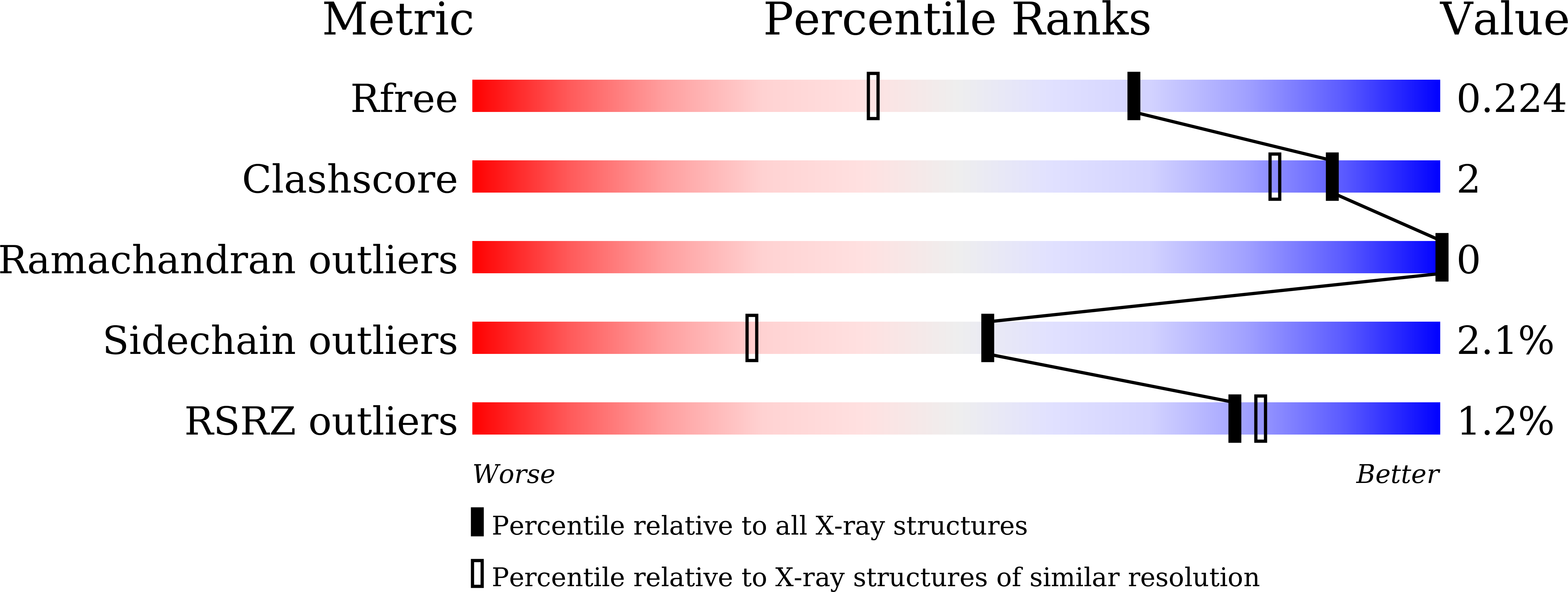
  • R-Value Free: 0.221 
  • R-Value Work: 0.182 
  • R-Value Observed: 0.184 

Starting Model: experimental
View more details

wwPDB Validation   3D Report Full Report


Ligand Structure Quality Assessment 


This is version 1.2 of the entry. See complete history


Literature

Protocatechuate hydroxylase is a novel group A flavoprotein monooxygenase with a unique substrate recognition mechanism.

Katsuki, N.Fukushima, R.Doi, Y.Masuo, S.Arakawa, T.Yamada, C.Fushinobu, S.Takaya, N.

(2023) J Biol Chem 300: 105508-105508

  • DOI: https://doi.org/10.1016/j.jbc.2023.105508
  • Primary Citation of Related Structures:  
    8JQO, 8JQP, 8JQQ

  • PubMed Abstract: 

    Para-hydroxybenzoate hydroxylase (PHBH) is a group A flavoprotein monooxygenase that hydroxylates p-hydroxybenzoate to protocatechuate (PCA). Despite intensive studies of Pseudomonas aeruginosa p-hydroxybenzoate hydroxylase (PaPobA), the catalytic reactions of extremely diverse putative PHBH isozymes remain unresolved. We analyzed the phylogenetic relationships of known and predicted PHBHs and identified eight divergent clades. Clade F contains a protein that lacks the critical amino acid residues required for PaPobA to generate PHBH activity. Among proteins in this clade, Xylophilus ampelinus PobA (XaPobA) preferred PCA as a substrate and is the first known natural PCA 5-hydroxylase (PCAH). Crystal structures and kinetic properties revealed similar mechanisms of substrate carboxy group recognition between XaPobA and PaPobA. The unique Ile75, Met72, Val199, Trp201, and Phe385 residues of XaPobA form the bottom of a hydrophobic cavity with a shape that complements the 3-and 4-hydroxy groups of PCA and its binding site configuration. An interaction between the δ-sulfur atom of Met210 and the aromatic ring of PCA is likely to stabilize XaPobA-PCA complexes. The 4-hydroxy group of PCA forms a hydrogen bond with the main chain carbonyl of Thr294. These modes of binding constitute a novel substrate recognition mechanism that PaPobA lacks. This mechanism characterizes XaPobA and sheds light on the diversity of catalytic mechanisms of PobA-type PHBHs and group A flavoprotein monooxygenases.


  • Organizational Affiliation

    Faculty of Life and Environmental Sciences, Microbiology Research Center for Sustainability, University of Tsukuba, Tsukuba, Ibaraki, Japan.


Macromolecules
Find similar proteins by:  (by identity cutoff)  |  3D Structure
Entity ID: 1
MoleculeChains Sequence LengthOrganismDetailsImage
4-hydroxybenzoate 3-monooxygenase (NAD(P)H)
A, B
411Xylophilus ampelinusMutation(s): 0 
Gene Names: praI
Entity Groups  
Sequence Clusters30% Identity50% Identity70% Identity90% Identity95% Identity100% Identity
Sequence Annotations
Expand
  • Reference Sequence
Small Molecules
Ligands 4 Unique
IDChains Name / Formula / InChI Key2D Diagram3D Interactions
FAD (Subject of Investigation/LOI)
Query on FAD

Download Ideal Coordinates CCD File 
C [auth A],
H [auth B]
FLAVIN-ADENINE DINUCLEOTIDE
C27 H33 N9 O15 P2
VWWQXMAJTJZDQX-UYBVJOGSSA-N
DHB (Subject of Investigation/LOI)
Query on DHB

Download Ideal Coordinates CCD File 
D [auth A],
E [auth A],
I [auth B],
J [auth B]
3,4-DIHYDROXYBENZOIC ACID
C7 H6 O4
YQUVCSBJEUQKSH-UHFFFAOYSA-N
GOL
Query on GOL

Download Ideal Coordinates CCD File 
F [auth A],
K [auth B]
GLYCEROL
C3 H8 O3
PEDCQBHIVMGVHV-UHFFFAOYSA-N
CA
Query on CA

Download Ideal Coordinates CCD File 
G [auth A],
L [auth B]
CALCIUM ION
Ca
BHPQYMZQTOCNFJ-UHFFFAOYSA-N
Experimental Data & Validation

Experimental Data

  • Method: X-RAY DIFFRACTION
  • Resolution: 1.65 Å
  • R-Value Free: 0.221 
  • R-Value Work: 0.182 
  • R-Value Observed: 0.184 
  • Space Group: P 1 21 1
Unit Cell:
Length ( Å )Angle ( ˚ )
a = 72.966α = 90
b = 65.074β = 107.55
c = 80.681γ = 90
Software Package:
Software NamePurpose
REFMACrefinement
PHENIXrefinement
XDSdata reduction
Aimlessdata scaling
MOLREPphasing

Structure Validation

View Full Validation Report


Ligand Structure Quality Assessment 


Entry History & Funding Information

Deposition Data


Funding OrganizationLocationGrant Number
Japan Society for the Promotion of Science (JSPS)JapanJP19H05679
Japan Society for the Promotion of Science (JSPS)JapanJP19H05687

Revision History  (Full details and data files)

  • Version 1.0: 2023-12-06
    Type: Initial release
  • Version 1.1: 2023-12-13
    Changes: Database references
  • Version 1.2: 2024-01-03
    Changes: Database references