8KI5

PhmA, a type I diterpene synthase without NST/DTE motif


Experimental Data Snapshot

  • Method: X-RAY DIFFRACTION
  • Resolution: 2.01 Å
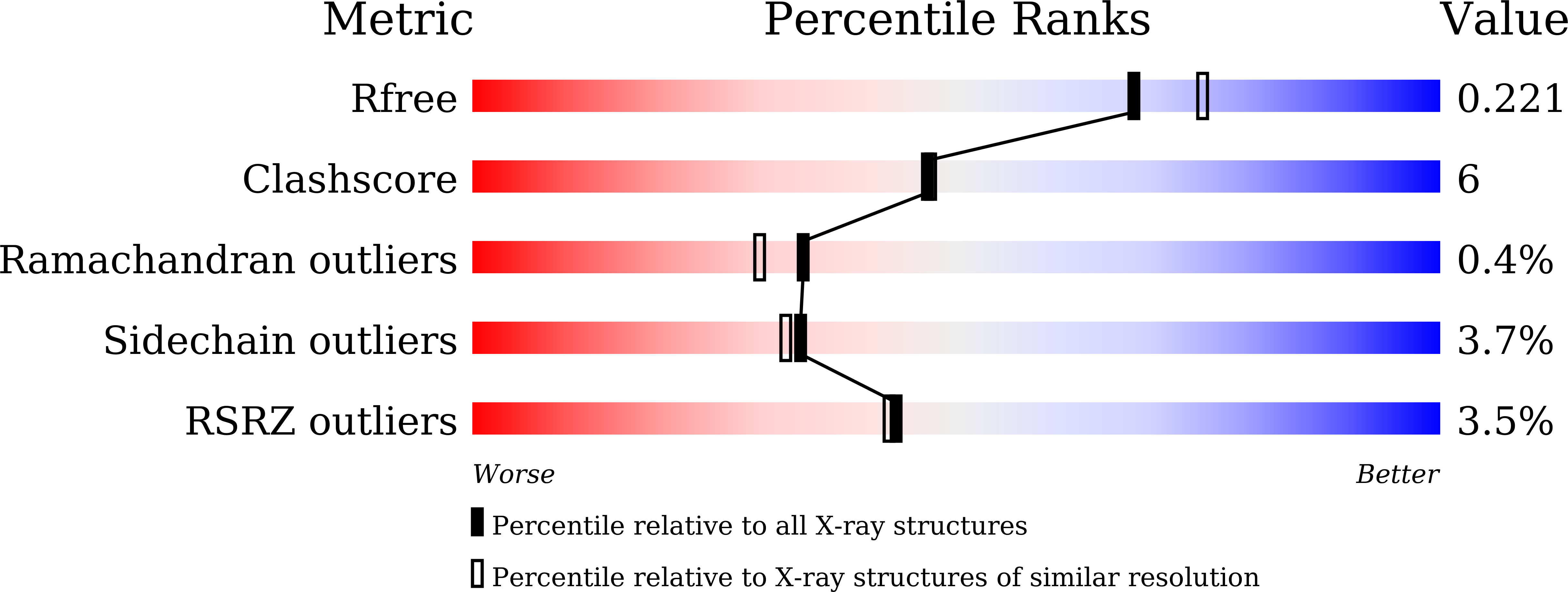
  • R-Value Free: 0.223 
  • R-Value Work: 0.182 
  • R-Value Observed: 0.186 

Starting Model: in silico
View more details

wwPDB Validation   3D Report Full Report


Ligand Structure Quality Assessment 


This is version 1.1 of the entry. See complete history


Literature

Biosynthesis of Phomactin Platelet Activating Factor Antagonist Requires a Two-Enzyme Cascade.

Zhang, L.Zhang, B.Zhu, A.Liu, S.H.Wu, R.Zhang, X.Xu, Z.Tan, R.X.Ge, H.M.

(2023) Angew Chem Int Ed Engl 62: e202312996-e202312996

  • DOI: https://doi.org/10.1002/anie.202312996
  • Primary Citation of Related Structures:  
    8KI5, 8KIH

  • PubMed Abstract: 

    Phomactin diterpenoids possess a unique bicyclo[9.3.1]pentadecane skeleton with multiple oxidative modifications, and are good platelet-activating factor (PAF) antagonists that can inhibit PAF-induced platelet aggregation. In this study, we identified the gene cluster (phm) responsible for the biosynthesis of phomactins from a marine fungus, Phoma sp. ATCC 74077. Despite the complexity of their structures, phomactin biosynthesis only requires two enzymes: a type I diterpene cyclase PhmA and a P450 monooxygenase PhmC. PhmA was found to catalyze the formation of the phomactatriene, while PhmC sequentially catalyzes the oxidation of multiple sites, leading to the generation of structurally diverse phomactins. The rearrangement mechanism of the diterpene scaffold was investigated through isotope labeling experiments. Additionally, we obtained the crystal complex of PhmA with its substrate analogue FGGPP and elucidated the novel metal-ion-binding mode and enzymatic mechanism of PhmA through site-directed mutagenesis. This study provides the first insight into the biosynthesis of phomactins, laying the foundation for the efficient production of phomactin natural products using synthetic biology approaches.


  • Organizational Affiliation

    State Key Laboratory of Pharmaceutical Biotechnology, Department of Neurology, Nanjing Drum Tower Hospital, School of Life Sciences, Chemistry and Biomedicine Innovation Center (ChemBIC), Nanjing University, Nanjing, 210023, China.


Macromolecules
Find similar proteins by:  (by identity cutoff)  |  3D Structure
Entity ID: 1
MoleculeChains Sequence LengthOrganismDetailsImage
PhmA332PhomaMutation(s): 0 
Entity Groups  
Sequence Clusters30% Identity50% Identity70% Identity90% Identity95% Identity100% Identity
Sequence Annotations
Expand
  • Reference Sequence
Small Molecules
Ligands 1 Unique
IDChains Name / Formula / InChI Key2D Diagram3D Interactions
FOH (Subject of Investigation/LOI)
Query on FOH

Download Ideal Coordinates CCD File 
B [auth A](2Z,6Z)-3,7,11-trimethyldodeca-2,6,10-trien-1-ol
C15 H26 O
CRDAMVZIKSXKFV-FBXUGWQNSA-N
Experimental Data & Validation

Experimental Data

  • Method: X-RAY DIFFRACTION
  • Resolution: 2.01 Å
  • R-Value Free: 0.223 
  • R-Value Work: 0.182 
  • R-Value Observed: 0.186 
  • Space Group: P 64
Unit Cell:
Length ( Å )Angle ( ˚ )
a = 105.9α = 90
b = 105.9β = 90
c = 54.38γ = 120
Software Package:
Software NamePurpose
PHENIXrefinement
XDSdata scaling
XDSdata reduction
PHENIXphasing

Structure Validation

View Full Validation Report


Ligand Structure Quality Assessment 


Entry History & Funding Information

Deposition Data


Funding OrganizationLocationGrant Number
National Natural Science Foundation of China (NSFC)China22277051, 22193071, 22107048, 22077062, 81991522, 81991524
Other governmentChinaBK20220123

Revision History  (Full details and data files)

  • Version 1.0: 2023-10-04
    Type: Initial release
  • Version 1.1: 2024-10-16
    Changes: Database references, Structure summary