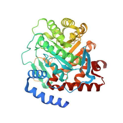
8RAK

Crystal structure of human Dihydroorotate Dehydrogenase in complex with the inhibitor 2-Hydroxy-N-(2-isopropyl-5-methyl-4-(pyridin-4-yloxy)phenyl)pyrazolo[1,5-a]pyridine-3-carboxamide


Experimental Data Snapshot

  • Method: X-RAY DIFFRACTION
  • Resolution: 1.85 Å
  • R-Value Free: 0.186 
  • R-Value Work: 0.164 
  • R-Value Observed: 0.166 

Starting Model: experimental
View more details

wwPDB Validation   3D Report Full Report


Ligand Structure Quality Assessment 


This is version 1.0 of the entry. See complete history


Literature

An alternative conformation of the N-terminal loop of human dihydroorotate dehydrogenase drives binding to a potent antiproliferative agent.

Alberti, M.Poli, G.Broggini, L.Sainas, S.Rizzi, M.Boschi, D.Ferraris, D.M.Martino, E.Ricagno, S.Tuccinardi, T.Lolli, M.L.Miggiano, R.

(2024) Acta Crystallogr D Struct Biol 80: 386-396

  • DOI: https://doi.org/10.1107/S2059798324004066
  • Primary Citation of Related Structures:  
    8RAK

  • PubMed Abstract: 

    Over the years, human dihydroorotate dehydrogenase (hDHODH), which is a key player in the de novo pyrimidine-biosynthesis pathway, has been targeted in the treatment of several conditions, including autoimmune disorders and acute myelogenous leukaemia, as well as in host-targeted antiviral therapy. A molecular exploration of its inhibitor-binding behaviours yielded promising candidates for innovative drug design. A detailed description of the enzymatic pharmacophore drove the decoration of well-established inhibitory scaffolds, thus gaining further in vitro and in vivo efficacy. In the present work, using X-ray crystallography, an atypical rearrangement was identified in the binding pose of a potent inhibitor characterized by a polar pyridine-based moiety (compound 18). The crystal structure shows that upon binding compound 18 the dynamics of a protein loop involved in a gating mechanism at the cofactor-binding site is modulated by the presence of three water molecules, thus fine-tuning the polarity/hydrophobicity of the binding pocket. These solvent molecules are engaged in the formation of a hydrogen-bond mesh in which one of them establishes a direct contact with the pyridine moiety of compound 18, thus paving the way for a reappraisal of the inhibition of hDHODH. Using an integrated approach, the thermodynamics of such a modulation is described by means of isothermal titration calorimetry coupled with molecular modelling. These structural insights will guide future drug design to obtain a finer K d /logD 7.4 balance and identify membrane-permeable molecules with a drug-like profile in terms of water solubility.


  • Organizational Affiliation

    Department of Pharmaceutical Sciences, University of Piemonte Orientale, Via G. Bovio 6, 28100 Novara, Italy.


Macromolecules
Find similar proteins by:  (by identity cutoff)  |  3D Structure
Entity ID: 1
MoleculeChains Sequence LengthOrganismDetailsImage
Dihydroorotate dehydrogenase (quinone), mitochondrial390Homo sapiensMutation(s): 0 
Gene Names: DHODH
EC: 1.3.5.2
UniProt & NIH Common Fund Data Resources
Find proteins for Q02127 (Homo sapiens)
Explore Q02127 
Go to UniProtKB:  Q02127
PHAROS:  Q02127
GTEx:  ENSG00000102967 
Entity Groups  
Sequence Clusters30% Identity50% Identity70% Identity90% Identity95% Identity100% Identity
UniProt GroupQ02127
Sequence Annotations
Expand
  • Reference Sequence
Small Molecules
Ligands 5 Unique
IDChains Name / Formula / InChI Key2D Diagram3D Interactions
FMN
Query on FMN

Download Ideal Coordinates CCD File 
B [auth A]FLAVIN MONONUCLEOTIDE
C17 H21 N4 O9 P
FVTCRASFADXXNN-SCRDCRAPSA-N
YMH (Subject of Investigation/LOI)
Query on YMH

Download Ideal Coordinates CCD File 
D [auth A]~{N}-(5-methyl-2-propan-2-yl-4-pyridin-4-yloxy-phenyl)-2-oxidanyl-pyrazolo[1,5-a]pyridine-3-carboxamide
C23 H22 N4 O3
IGBISIGCTJJXOY-UHFFFAOYSA-N
ORO
Query on ORO

Download Ideal Coordinates CCD File 
C [auth A]OROTIC ACID
C5 H4 N2 O4
PXQPEWDEAKTCGB-UHFFFAOYSA-N
SO4
Query on SO4

Download Ideal Coordinates CCD File 
J [auth A],
K [auth A]
SULFATE ION
O4 S
QAOWNCQODCNURD-UHFFFAOYSA-L
ACT
Query on ACT

Download Ideal Coordinates CCD File 
E [auth A],
F [auth A],
G [auth A],
H [auth A],
I [auth A]
ACETATE ION
C2 H3 O2
QTBSBXVTEAMEQO-UHFFFAOYSA-M
Experimental Data & Validation

Experimental Data

  • Method: X-RAY DIFFRACTION
  • Resolution: 1.85 Å
  • R-Value Free: 0.186 
  • R-Value Work: 0.164 
  • R-Value Observed: 0.166 
  • Space Group: P 32 2 1
Unit Cell:
Length ( Å )Angle ( ˚ )
a = 91.474α = 90
b = 91.474β = 90
c = 124.032γ = 120
Software Package:
Software NamePurpose
PHENIXrefinement
PHENIXrefinement
XDSdata reduction
SCALAdata scaling
PHASERphasing

Structure Validation

View Full Validation Report


Ligand Structure Quality Assessment 


Entry History & Funding Information

Deposition Data


Funding OrganizationLocationGrant Number
Other governmentItalyDSF-FAR 2017

Revision History  (Full details and data files)

  • Version 1.0: 2024-06-19
    Type: Initial release