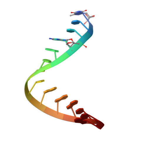
Insight into the structures of unusual base pairs in RNA complexes containing a primer/template/adenosine ligand.
Dantsu, Y., Zhang, Y., Zhang, W.(2023) RSC Chem Biol 4: 942-951
- PubMed: 37920395
- DOI: https://doi.org/10.1039/d3cb00137g
- Primary Citation of Related Structures:
8SWG, 8SWO, 8SX5, 8SX6, 8SXL, 8SY1 - PubMed Abstract:
In the prebiotic RNA world, the self-replication of RNA without enzymes can be achieved through the utilization of 2-aminoimidazole activated nucleotides as efficient substrates. The mechanism of RNA nonenzymatic polymerization has been extensively investigated biophysically and structurally by using the model of an RNA primer/template complex which is bound by the imidazolium-bridged or triphosphate-bridged diguanosine intermediate. However, beyond the realm of the guanosine substrate, the structural insight into how alternative activated nucleotides bind and interact with the RNA primer/template complex remains unexplored, which is important for understanding the low reactivity of adenosine and uridine substrates in RNA primer extension, as well as its relationship with the structures. Here we use crystallography as a method and determine a series of high-resolution structures of RNA primer/template complexes bound by ApppG, a close analog of the dinucleotide intermediate containing adenosine and guanosine. The structures show that ApppG ligands bind to the RNA template through both Watson-Crick and noncanonical base pairs, with the primer 3'-OH group far from the adjacent phosphorus atom of the incoming substrate. The structures indicate that when adenosine is included in the imidazolium-bridged intermediate, the complexes are likely preorganized in a suboptimal conformation, making it difficult for the primer to in-line attack the substrate. Moreover, by co-crystallizing the RNA primer/template with chemically activated adenosine and guanosine monomers, we successfully observe the slow formation of the imidazolium-bridged intermediate (Ap-AI-pG) and the preorganized structure for RNA primer extension. Overall, our studies offer a structural explanation for the slow rate of RNA primer extension when using adenosine-5'-phosphoro-2-aminoimidazolide as a substrate during nonenzymatic polymerization.
Organizational Affiliation:
Department of Biochemistry and Molecular Biology, Indiana University School of Medicine Indianapolis IN 46202 USA [email protected].


