8TE3

Crystal structure of the methyltransferase domain of R882C/R676K DNMT3A homotetramer


Experimental Data Snapshot

  • Method: X-RAY DIFFRACTION
  • Resolution: 3.20 Å
  • R-Value Free: 0.241 
  • R-Value Work: 0.207 
  • R-Value Observed: 0.208 

wwPDB Validation   3D Report Full Report


Ligand Structure Quality Assessment 


This is version 1.1 of the entry. See complete history


Literature

Structure-guided functional suppression of AML-associated DNMT3A hotspot mutations.

Lu, J.Guo, Y.Yin, J.Chen, J.Wang, Y.Wang, G.G.Song, J.

(2024) Nat Commun 15: 3111-3111

  • DOI: https://doi.org/10.1038/s41467-024-47398-y
  • Primary Citation of Related Structures:  
    8TDR, 8TE1, 8TE3, 8TE4

  • PubMed Abstract: 

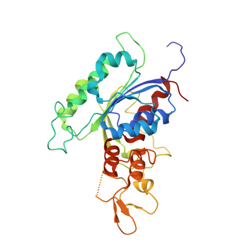
    DNA methyltransferases DNMT3A- and DNMT3B-mediated DNA methylation critically regulate epigenomic and transcriptomic patterning during development. The hotspot DNMT3A mutations at the site of Arg822 (R882) promote polymerization, leading to aberrant DNA methylation that may contribute to the pathogenesis of acute myeloid leukemia (AML). However, the molecular basis underlying the mutation-induced functional misregulation of DNMT3A remains unclear. Here, we report the crystal structures of the DNMT3A methyltransferase domain, revealing a molecular basis for its oligomerization behavior distinct to DNMT3B, and the enhanced intermolecular contacts caused by the R882H or R882C mutation. Our biochemical, cellular, and genomic DNA methylation analyses demonstrate that introducing the DNMT3B-converting mutations inhibits the R882H-/R882C-triggered DNMT3A polymerization and enhances substrate access, thereby eliminating the dominant-negative effect of the DNMT3A R882 mutations in cells. Together, this study provides mechanistic insights into DNMT3A R882 mutations-triggered aberrant oligomerization and DNA hypomethylation in AML, with important implications in cancer therapy.


  • Organizational Affiliation

    Department of Biochemistry, University of California, Riverside, CA, USA.


Macromolecules
Find similar proteins by:  (by identity cutoff)  |  3D Structure
Entity ID: 1
MoleculeChains Sequence LengthOrganismDetailsImage
DNA (cytosine-5)-methyltransferase 3A287Homo sapiensMutation(s): 2 
Gene Names: DNMT3A
EC: 2.1.1.37 (PDB Primary Data), 2.1.1 (PDB Primary Data)
UniProt & NIH Common Fund Data Resources
Find proteins for Q9Y6K1 (Homo sapiens)
Explore Q9Y6K1 
Go to UniProtKB:  Q9Y6K1
PHAROS:  Q9Y6K1
GTEx:  ENSG00000119772 
Entity Groups  
Sequence Clusters30% Identity50% Identity70% Identity90% Identity95% Identity100% Identity
UniProt GroupQ9Y6K1
Sequence Annotations
Expand
  • Reference Sequence
Experimental Data & Validation

Experimental Data

  • Method: X-RAY DIFFRACTION
  • Resolution: 3.20 Å
  • R-Value Free: 0.241 
  • R-Value Work: 0.207 
  • R-Value Observed: 0.208 
  • Space Group: P 3
Unit Cell:
Length ( Å )Angle ( ˚ )
a = 177.298α = 90
b = 177.298β = 90
c = 110.736γ = 120
Software Package:
Software NamePurpose
PHENIXrefinement
HKL-2000data scaling
PDB_EXTRACTdata extraction
HKL-3000data reduction
PHENIXphasing

Structure Validation

View Full Validation Report


Ligand Structure Quality Assessment 


Entry History & Funding Information

Deposition Data


Funding OrganizationLocationGrant Number
National Institutes of Health/National Cancer Institute (NIH/NCI)United States--

Revision History  (Full details and data files)

  • Version 1.0: 2024-03-13
    Type: Initial release
  • Version 1.1: 2024-09-25
    Changes: Data collection, Database references