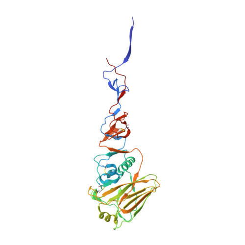
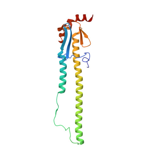
8TJ4

CRYSTAL STRUCTURE OF THE A/Bangkok/1/1979(H3N2) INFLUENZA VIRUS HEMAGGLUTININ WITH HUMAN RECEPTOR ANALOG 6'-SLNLN


Experimental Data Snapshot

  • Method: X-RAY DIFFRACTION
  • Resolution: 1.95 Å
  • R-Value Free: 0.223 
  • R-Value Work: 0.189 
  • R-Value Observed: 0.191 

Starting Model: experimental
View more details

wwPDB Validation   3D Report Full Report


Ligand Structure Quality Assessment 


This is version 1.2 of the entry. See complete history


Literature

Evolution of human H3N2 influenza virus receptor specificity has substantially expanded the receptor-binding domain site.

Thompson, A.J.Wu, N.C.Canales, A.Kikuchi, C.Zhu, X.de Toro, B.F.Canada, F.J.Worth, C.Wang, S.McBride, R.Peng, W.Nycholat, C.M.Jimenez-Barbero, J.Wilson, I.A.Paulson, J.C.

(2024) Cell Host Microbe 32: 261

  • DOI: https://doi.org/10.1016/j.chom.2024.01.003
  • Primary Citation of Related Structures:  
    8TJ4, 8TJ6, 8TJ7, 8TJ8, 8TJ9, 8TJA, 8TJB

  • PubMed Abstract: 

    Hemagglutinins (HAs) from human influenza viruses descend from avian progenitors that bind α2-3-linked sialosides and must adapt to glycans with α2-6-linked sialic acids on human airway cells to transmit within the human population. Since their introduction during the 1968 pandemic, H3N2 viruses have evolved over the past five decades to preferentially recognize human α2-6-sialoside receptors that are elongated through addition of poly-LacNAc. We show that more recent H3N2 viruses now make increasingly complex interactions with elongated receptors while continuously selecting for strains maintaining this phenotype. This change in receptor engagement is accompanied by an extension of the traditional receptor-binding site to include residues in key antigenic sites on the surface of HA trimers. These results help explain the propensity for selection of antigenic variants, leading to vaccine mismatching, when H3N2 viruses are propagated in chicken eggs or cells that do not contain such receptors.


  • Organizational Affiliation

    Department of Molecular Medicine, The Scripps Research Institute, La Jolla, CA 92037, USA.


Macromolecules
Find similar proteins by:  (by identity cutoff)  |  3D Structure
Entity ID: 1
MoleculeChains Sequence LengthOrganismDetailsImage
Hemagglutinin HA1 chain
A, C, E, G
323Influenza A virus (A/Bangkok/1/1979(H3N2))Mutation(s): 0 
Gene Names: HA
UniProt
Find proteins for P03441 (Influenza A virus (strain A/Bangkok/1/1979 H3N2))
Explore P03441 
Go to UniProtKB:  P03441
Entity Groups  
Sequence Clusters30% Identity50% Identity70% Identity90% Identity95% Identity100% Identity
UniProt GroupP03441
Glycosylation
Glycosylation Sites: 5
Sequence Annotations
Expand
  • Reference Sequence
Find similar proteins by:  (by identity cutoff)  |  3D Structure
Entity ID: 2
MoleculeChains Sequence LengthOrganismDetailsImage
Hemagglutinin HA2 chain
B, D, F, H
174Influenza A virus (A/Bangkok/1/1979(H3N2))Mutation(s): 0 
Gene Names: HA
UniProt
Find proteins for P03441 (Influenza A virus (strain A/Bangkok/1/1979 H3N2))
Explore P03441 
Go to UniProtKB:  P03441
Entity Groups  
Sequence Clusters30% Identity50% Identity70% Identity90% Identity95% Identity100% Identity
UniProt GroupP03441
Glycosylation
Glycosylation Sites: 1
Sequence Annotations
Expand
  • Reference Sequence
Oligosaccharides

Help

Entity ID: 3
MoleculeChains Length2D Diagram Glycosylation3D Interactions
2-acetamido-2-deoxy-beta-D-glucopyranose-(1-4)-2-acetamido-2-deoxy-beta-D-glucopyranose
I, L, O, S, T
I, L, O, S, T, U, V, W, X
2N-Glycosylation
Glycosylation Resources
GlyTouCan:  G42666HT
GlyCosmos:  G42666HT
GlyGen:  G42666HT
Entity ID: 4
MoleculeChains Length2D Diagram Glycosylation3D Interactions
beta-D-mannopyranose-(1-4)-2-acetamido-2-deoxy-beta-D-glucopyranose-(1-4)-2-acetamido-2-deoxy-beta-D-glucopyranose
J, M, N
3N-Glycosylation
Glycosylation Resources
GlyTouCan:  G15407YE
GlyCosmos:  G15407YE
GlyGen:  G15407YE
Entity ID: 5
MoleculeChains Length2D Diagram Glycosylation3D Interactions
alpha-D-mannopyranose-(1-3)-[alpha-D-mannopyranose-(1-6)]beta-D-mannopyranose-(1-4)-2-acetamido-2-deoxy-beta-D-glucopyranose-(1-4)-2-acetamido-2-deoxy-beta-D-glucopyranose
K, Q, R
5N-Glycosylation
Glycosylation Resources
GlyTouCan:  G22768VO
GlyCosmos:  G22768VO
GlyGen:  G22768VO
Entity ID: 6
MoleculeChains Length2D Diagram Glycosylation3D Interactions
alpha-L-fucopyranose-(1-3)-[2-acetamido-2-deoxy-beta-D-glucopyranose-(1-4)][alpha-L-fucopyranose-(1-6)]2-acetamido-2-deoxy-beta-D-glucopyranose
P, Y
4N-Glycosylation
Glycosylation Resources
GlyTouCan:  G63564LA
GlyCosmos:  G63564LA
Entity ID: 7
MoleculeChains Length2D Diagram Glycosylation3D Interactions
alpha-D-mannopyranose-(1-3)-alpha-D-mannopyranose-(1-6)-[alpha-D-mannopyranose-(1-3)]beta-D-mannopyranose-(1-4)-2-acetamido-2-deoxy-beta-D-glucopyranose-(1-4)-2-acetamido-2-deoxy-beta-D-glucopyranose
Z
6N-Glycosylation
Glycosylation Resources
GlyTouCan:  G09724ZC
GlyCosmos:  G09724ZC
GlyGen:  G09724ZC
Entity ID: 8
MoleculeChains Length2D Diagram Glycosylation3D Interactions
N-acetyl-alpha-neuraminic acid-(2-6)-beta-D-galactopyranose-(1-4)-2-acetamido-2-deoxy-beta-D-glucopyranoseAA [auth a],
CA [auth c]
3N/A
Glycosylation Resources
GlyTouCan:  G73578JC
GlyCosmos:  G73578JC
GlyGen:  G73578JC
Entity ID: 9
MoleculeChains Length2D Diagram Glycosylation3D Interactions
N-acetyl-alpha-neuraminic acid-(2-6)-beta-D-galactopyranoseBA [auth b],
DA [auth d]
2N/A
Glycosylation Resources
GlyTouCan:  G63069TR
GlyCosmos:  G63069TR
GlyGen:  G63069TR
Small Molecules
Ligands 4 Unique
IDChains Name / Formula / InChI Key2D Diagram3D Interactions
NAG (Subject of Investigation/LOI)
Query on NAG

Download Ideal Coordinates CCD File 
EA [auth A]
FA [auth B]
IA [auth D]
JA [auth E]
LA [auth F]
EA [auth A],
FA [auth B],
IA [auth D],
JA [auth E],
LA [auth F],
PA [auth G],
QA [auth H]
2-acetamido-2-deoxy-beta-D-glucopyranose
C8 H15 N O6
OVRNDRQMDRJTHS-FMDGEEDCSA-N
PG4
Query on PG4

Download Ideal Coordinates CCD File 
GA [auth B],
MA [auth F]
TETRAETHYLENE GLYCOL
C8 H18 O5
UWHCKJMYHZGTIT-UHFFFAOYSA-N
PG0
Query on PG0

Download Ideal Coordinates CCD File 
HA [auth B],
RA [auth H]
2-(2-METHOXYETHOXY)ETHANOL
C5 H12 O3
SBASXUCJHJRPEV-UHFFFAOYSA-N
PEG
Query on PEG

Download Ideal Coordinates CCD File 
KA [auth E],
NA [auth F],
OA [auth F]
DI(HYDROXYETHYL)ETHER
C4 H10 O3
MTHSVFCYNBDYFN-UHFFFAOYSA-N
Experimental Data & Validation

Experimental Data

  • Method: X-RAY DIFFRACTION
  • Resolution: 1.95 Å
  • R-Value Free: 0.223 
  • R-Value Work: 0.189 
  • R-Value Observed: 0.191 
  • Space Group: H 3
Unit Cell:
Length ( Å )Angle ( ˚ )
a = 100.655α = 90
b = 100.655β = 90
c = 687.903γ = 120
Software Package:
Software NamePurpose
PHENIXrefinement
HKL-2000data reduction
HKL-2000data scaling
PHASERphasing

Structure Validation

View Full Validation Report


Ligand Structure Quality Assessment 


Entry History & Funding Information

Deposition Data


Funding OrganizationLocationGrant Number
National Institutes of Health/National Institute Of Allergy and Infectious Diseases (NIH/NIAID)United States--
Bill & Melinda Gates FoundationUnited States--

Revision History  (Full details and data files)

  • Version 1.0: 2024-02-14
    Type: Initial release
  • Version 1.1: 2024-02-28
    Changes: Database references
  • Version 1.2: 2024-11-13
    Changes: Structure summary