9C1A

Crystal structure of GDP-bound human M-RAS protein in crystal form I


Experimental Data Snapshot

  • Method: X-RAY DIFFRACTION
  • Resolution: 1.96 Å
  • R-Value Free: 0.200 
  • R-Value Work: 0.172 
  • R-Value Observed: 0.175 

Starting Model: experimental
View more details

wwPDB Validation   3D Report Full Report


Ligand Structure Quality Assessment 


This is version 1.1 of the entry. See complete history


Literature

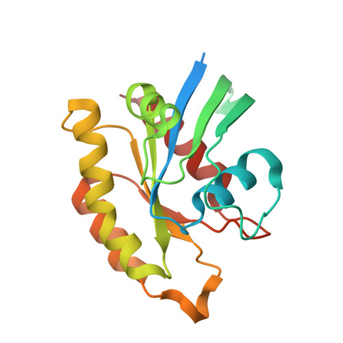
Crystal structure of the GDP-bound human M-RAS protein in two crystal forms.

Bester, S.M.Abrahamsen, R.Rodrigues Samora, L.Wu, W.I.Mou, T.C.

(2024) Acta Crystallogr F Struct Biol Commun 80: 220-227

  • DOI: https://doi.org/10.1107/S2053230X24007969
  • Primary Citation of Related Structures:  
    9C1A, 9C1B

  • PubMed Abstract: 

    M-RAS plays a crucial role in the RAF-MEK signaling pathway. When activated by GTP, M-RAS forms a complex with SHOC2 and PP1C, initiating downstream RAF-MEK signal transduction. In this study, the crystal structure of the GDP-bound human M-RAS protein is presented with two forms of crystal packing. Both the full-length and truncated human M-RAS structures aligned well with the high-confidence section of the AlphaFold2-predicted structure with low r.m.s.d., except for the Switch regions. Despite high sequence similarity to the available mouse M-RAS structure, the full-length human M-RAS structure exhibits unique crystal packing. This inactive human M-RAS structure could offer novel insights for the design of selective compounds targeting M-RAS.


  • Organizational Affiliation

    Pfizer Boulder Research and Development, 3200 Walnut Street, Boulder, CO 80301, USA.


Macromolecules
Find similar proteins by:  (by identity cutoff)  |  3D Structure
Entity ID: 1
MoleculeChains Sequence LengthOrganismDetailsImage
Ras-related protein M-Ras193Homo sapiensMutation(s): 0 
Gene Names: MRASRRAS3
EC: 3.6.5.2
UniProt & NIH Common Fund Data Resources
Find proteins for O14807 (Homo sapiens)
Explore O14807 
Go to UniProtKB:  O14807
PHAROS:  O14807
GTEx:  ENSG00000158186 
Entity Groups  
Sequence Clusters30% Identity50% Identity70% Identity90% Identity95% Identity100% Identity
UniProt GroupO14807
Sequence Annotations
Expand
  • Reference Sequence
Experimental Data & Validation

Experimental Data

  • Method: X-RAY DIFFRACTION
  • Resolution: 1.96 Å
  • R-Value Free: 0.200 
  • R-Value Work: 0.172 
  • R-Value Observed: 0.175 
  • Space Group: P 6 2 2
Unit Cell:
Length ( Å )Angle ( ˚ )
a = 108.653α = 90
b = 108.653β = 90
c = 65.759γ = 120
Software Package:
Software NamePurpose
PHENIXrefinement
PHENIXrefinement
autoPROCdata reduction
autoPROCdata scaling
PHASERphasing

Structure Validation

View Full Validation Report


Ligand Structure Quality Assessment 


Entry History & Funding Information

Deposition Data


Funding OrganizationLocationGrant Number
Other private--

Revision History  (Full details and data files)

  • Version 1.0: 2024-09-11
    Type: Initial release
  • Version 1.1: 2024-09-18
    Changes: Database references