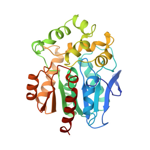
Haloalkane dehalogenases: structure of a Rhodococcus enzyme.
Newman, J., Peat, T.S., Richard, R., Kan, L., Swanson, P.E., Affholter, J.A., Holmes, I.H., Schindler, J.F., Unkefer, C.J., Terwilliger, T.C.(1999) Biochemistry 38: 16105-16114
- PubMed: 10587433
- DOI: https://doi.org/10.1021/bi9913855
- Primary Citation of Related Structures:
1BN6, 1BN7, 1CQW - PubMed Abstract:
The hydrolytic haloalkane dehalogenases are promising bioremediation and biocatalytic agents. Two general classes of dehalogenases have been reported from Xanthobacter and Rhodococcus. While these enzymes share 30% amino acid sequence identity, they have significantly different substrate specificities and halide-binding properties. We report the 1.5 A resolution crystal structure of the Rhodococcus dehalogenase at pH 5.5, pH 7.0, and pH 5.5 in the presence of NaI. The Rhodococcus and Xanthobacter enzymes have significant structural homology in the alpha/beta hydrolase core, but differ considerably in the cap domain. Consistent with its broad specificity for primary, secondary, and cyclic haloalkanes, the Rhodococcus enzyme has a substantially larger active site cavity. Significantly, the Rhodococcus dehalogenase has a different catalytic triad topology than the Xanthobacter enzyme. In the Xanthobacter dehalogenase, the third carboxylate functionality in the triad is provided by D260, which is positioned on the loop between beta7 and the penultimate helix. The carboxylate functionality in the Rhodococcus catalytic triad is donated from E141. A model of the enzyme cocrystallized with sodium iodide shows two iodide binding sites; one that defines the normal substrate and product-binding site and a second within the active site region. In the substrate and product complexes, the halogen binds to the Xanthobacter enzyme via hydrogen bonds with the N(eta)H of both W125 and W175. The Rhodococcusenzyme does not have a tryptophan analogous to W175. Instead, bound halide is stabilized with hydrogen bonds to the N(eta)H of W118 and to N(delta)H of N52. It appears that when cocrystallized with NaI the Rhodococcus enzyme has a rare stable S-I covalent bond to S(gamma) of C187.
Organizational Affiliation:
Life Sciences Division, Los Alamos National Laboratory, New Mexico. [email protected]


