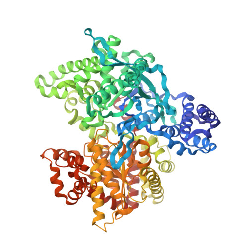
1E4O

Phosphorylase recognition and phosphorolysis of its oligosaccharide substrate: answers to a long outstanding question


Experimental Data Snapshot

  • Method: X-RAY DIFFRACTION
  • Resolution: 2.90 Å
  • R-Value Free: 0.306 
  • R-Value Work: 0.245 
  • R-Value Observed: 0.245 

wwPDB Validation   3D Report Full Report


This is version 2.0 of the entry. See complete history


Literature

Phosphorylase Recognition and Phosphorylysis of its Oligosaccharide Substrate: Answers to a Long Outstanding Question

Watson, K.A.Mccleverty, C.Geremia, S.Cottaz, S.Driguez, H.Johnson, L.N.

(1999) EMBO J 18: 4619

  • DOI: https://doi.org/10.1093/emboj/18.17.4619
  • Primary Citation of Related Structures:  
    1E4O, 1QM5

  • PubMed Abstract: 

    Phosphorylases are key enzymes of carbohydrate metabolism. Structural studies have provided explanations for almost all features of control and substrate recognition of phosphorylase but one question remains unanswered. How does phosphorylase recognize and cleave an oligosaccharide substrate? To answer this question we turned to the Escherichia coli maltodextrin phosphorylase (MalP), a non-regulatory phosphorylase that shares similar kinetic and catalytic properties with the mammalian glycogen phosphorylase. The crystal structures of three MalP-oligosaccharide complexes are reported: the binary complex of MalP with the natural substrate, maltopentaose (G5); the binary complex with the thio-oligosaccharide, 4-S-alpha-D-glucopyranosyl-4-thiomaltotetraose (GSG4), both at 2.9 A resolution; and the 2.1 A resolution ternary complex of MalP with thio-oligosaccharide and phosphate (GSG4-P). The results show a pentasaccharide bound across the catalytic site of MalP with sugars occupying sub-sites -1 to +4. Binding of GSG4 is identical to the natural pentasaccharide, indicating that the inactive thio compound is a close mimic of the natural substrate. The ternary MalP-GSG4-P complex shows the phosphate group poised to attack the glycosidic bond and promote phosphorolysis. In all three complexes the pentasaccharide exhibits an altered conformation across sub-sites -1 and +1, the site of catalysis, from the preferred conformation for alpha(1-4)-linked glucosyl polymers.


  • Organizational Affiliation

    Laboratory of Molecular Biophysics, Department of Biochemistry, University of Oxford, South Parks Road, Oxford OX1 3QU, UK. [email protected]


Macromolecules
Find similar proteins by:  (by identity cutoff)  |  3D Structure
Entity ID: 1
MoleculeChains Sequence LengthOrganismDetailsImage
MALTODEXTRIN PHOSPHORYLASE
A, B
796Escherichia coliMutation(s): 0 
EC: 2.4.1.1
UniProt
Find proteins for P00490 (Escherichia coli (strain K12))
Explore P00490 
Go to UniProtKB:  P00490
Entity Groups  
Sequence Clusters30% Identity50% Identity70% Identity90% Identity95% Identity100% Identity
UniProt GroupP00490
Sequence Annotations
Expand
  • Reference Sequence
Oligosaccharides

Help

Entity ID: 2
MoleculeChains Length2D Diagram Glycosylation3D Interactions
alpha-D-glucopyranose-(1-4)-alpha-D-glucopyranose-(1-4)-alpha-D-glucopyranose-(1-4)-alpha-D-glucopyranose-(1-4)-beta-D-glucopyranose
C, D
5N/A
Glycosylation Resources
GlyTouCan:  G43441FW
GlyCosmos:  G43441FW
Small Molecules
Ligands 1 Unique
IDChains Name / Formula / InChI Key2D Diagram3D Interactions
PLP
Query on PLP

Download Ideal Coordinates CCD File 
E [auth A],
F [auth B]
PYRIDOXAL-5'-PHOSPHATE
C8 H10 N O6 P
NGVDGCNFYWLIFO-UHFFFAOYSA-N
Experimental Data & Validation

Experimental Data

  • Method: X-RAY DIFFRACTION
  • Resolution: 2.90 Å
  • R-Value Free: 0.306 
  • R-Value Work: 0.245 
  • R-Value Observed: 0.245 
  • Space Group: P 21 21 21
Unit Cell:
Length ( Å )Angle ( ˚ )
a = 74.45α = 90
b = 105.69β = 90
c = 214.91γ = 90
Software Package:
Software NamePurpose
X-PLORrefinement
MOSFLMdata reduction
SCALAdata scaling
AMoREphasing

Structure Validation

View Full Validation Report


Entry History 

Deposition Data

Revision History  (Full details and data files)

  • Version 1.0: 2000-08-16
    Type: Initial release
  • Version 1.1: 2011-08-31
    Changes: Atomic model, Derived calculations, Non-polymer description, Other, Structure summary, Version format compliance
  • Version 1.2: 2012-05-30
    Changes: Other
  • Version 1.3: 2019-05-08
    Changes: Data collection, Derived calculations, Experimental preparation, Other
  • Version 2.0: 2020-07-29
    Type: Remediation
    Reason: Carbohydrate remediation
    Changes: Atomic model, Data collection, Derived calculations, Structure summary