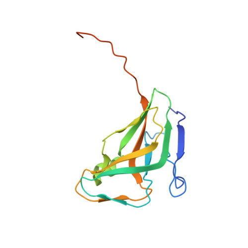
Atomic resolution structure of Escherichia coli dUTPase determined ab initio.
Gonzalez, A., Larsson, G., Persson, R., Cedergren-Zeppezauer, E.(2001) Acta Crystallogr D Biol Crystallogr 57: 767-774
- PubMed: 11375495
- DOI: https://doi.org/10.1107/s0907444901004255
- Primary Citation of Related Structures:
1EU5, 1EUW - PubMed Abstract:
Cryocooled crystals of a mercury complex of Escherichia coli dUTPase diffract to atomic resolution. Data to 1.05 A resolution were collected from a derivative crystal and the structure model was derived from a Fourier map with phases calculated from the coordinates of the Hg atom (one site per subunit of the trimeric enzyme) using the program ARP/wARP. After refinement with anisotropic temperature factors a highly accurate model of the bacterial dUTPase was obtained. Data to 1.45 A from a native crystal were also collected and the 100 K structures were compared. Inspection of the refined models reveals that a large part of the dUTPase remains rather mobile upon freezing, with 14% of the main chain being totally disordered and with numerous side chains containing disordered atoms in multiple discrete conformations. A large number of those residues surround the active-site cavity. Two glycerol molecules (the cryosolvent) occupy the deoxyribose-binding site. Comparison between the native enzyme and the mercury complex shows that the active site is not adversely affected by the binding of mercury. An unexpected effect seems to be a stabilization of the crystal lattice by means of long-range interactions, making derivatization a potentially useful tool for further studies of inhibitor-substrate-analogue complexes of this protein at very high resolution.
Organizational Affiliation:
European Molecular Biology Laboratory (EMBL), Hamburg Outstation, Notkestrasse 85, 22603 Hamburg, Germany. [email protected]


