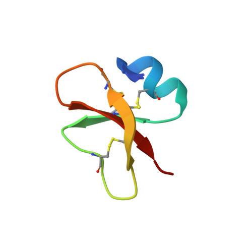
The structure of human beta-defensin-2 shows evidence of higher order oligomerization.
Hoover, D.M., Rajashankar, K.R., Blumenthal, R., Puri, A., Oppenheim, J.J., Chertov, O., Lubkowski, J.(2000) J Biol Chem 275: 32911-32918
- PubMed: 10906336
- DOI: https://doi.org/10.1074/jbc.M006098200
- Primary Citation of Related Structures:
1FD3, 1FD4 - PubMed Abstract:
Defensins are small cationic peptides that are crucial components of innate immunity, serving as both antimicrobial agents and chemoattractant molecules. The specific mechanism of antimicrobial activity involves permeabilization of bacterial membranes. It has been postulated that individual monomers oligomerize to form a pore through anionic membranes, although the evidence is only indirect. Here, we report two high resolution x-ray structures of human beta-defensin-2 (hBD2). The phases were experimentally determined by the multiwavelength anomalous diffraction method, utilizing a novel, rapid method of derivatization with halide ions. Although the shape and charge distribution of the monomer are similar to those of other defensins, an additional alpha-helical region makes this protein topologically distinct from the mammalian alpha- and beta-defensin structures reported previously. hBD2 forms dimers topologically distinct from that of human neutrophil peptide-3. The quaternary octameric arrangement of hBD2 is conserved in two crystal forms. These structures provide the first detailed description of dimerization of beta-defensins, and we postulate that the mode of dimerization of hBD2 is representative of other beta-defensins. The structural and electrostatic properties of the hBD2 octamer support an electrostatic charge-based mechanism of membrane permeabilization by beta-defensins, rather than a mechanism based on formation of bilayer-spanning pores.
Organizational Affiliation:
Macromolecular Crystallography Laboratory, Program in Structural Biology, NCI-Frederick Cancer Research and Development Center, National Institutes of Health, Frederick, Maryland 21702, USA.


