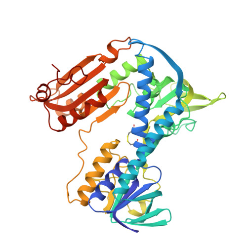
1GER

THE STRUCTURE OF GLUTATHIONE REDUCTASE FROM ESCHERICHIA COLI AT 1.86 ANGSTROMS RESOLUTION: COMPARISON WITH THE ENZYME FROM HUMAN ERYTHROCYTES


Experimental Data Snapshot

  • Method: X-RAY DIFFRACTION
  • Resolution: 1.86 Å
  • R-Value Work: 0.168 
  • R-Value Observed: 0.168 

wwPDB Validation   3D Report Full Report


Ligand Structure Quality Assessment 


This is version 1.4 of the entry. See complete history


Literature

Structure of glutathione reductase from Escherichia coli at 1.86 A resolution: comparison with the enzyme from human erythrocytes.

Mittl, P.R.Schulz, G.E.

(1994) Protein Sci 3: 799-809

  • DOI: https://doi.org/10.1002/pro.5560030509
  • Primary Citation of Related Structures:  
    1GER

  • PubMed Abstract: 

    The crystal structure of the dimeric flavoenzyme glutathione reductase from Escherichia coli was determined and refined to an R-factor of 16.8% at 1.86 A resolution. The molecular 2-fold axis of the dimer is local but very close to a possible crystallographic 2-fold axis; the slight asymmetry could be rationalized from the packing contacts. The 2 crystallographically independent subunits of the dimer are virtually identical, yielding no structural clue on possible cooperativity. The structure was compared with the well-known structure of the homologous enzyme from human erythrocytes with 52% sequence identity. Significant differences were found at the dimer interface, where the human enzyme has a disulfide bridge, whereas the E. coli enzyme has an antiparallel beta-sheet connecting the subunits. The differences at the glutathione binding site and in particular a deformation caused by a Leu-Ile exchange indicate why the E. coli enzyme accepts trypanothione much better than the human enzyme. The reported structure provides a frame for explaining numerous published engineering results in detail and for guiding further ones.


  • Organizational Affiliation

    Institut für Organische Chemie und Biochemie, Albert-Ludwigs-Universität, Freiburg, Germany.


Macromolecules
Find similar proteins by:  (by identity cutoff)  |  3D Structure
Entity ID: 1
MoleculeChains Sequence LengthOrganismDetailsImage
GLUTATHIONE REDUCTASE
A, B
450Escherichia coliMutation(s): 0 
EC: 1.6.4.2 (PDB Primary Data), 1.8.1.7 (UniProt)
UniProt
Find proteins for P06715 (Escherichia coli (strain K12))
Explore P06715 
Go to UniProtKB:  P06715
Entity Groups  
Sequence Clusters30% Identity50% Identity70% Identity90% Identity95% Identity100% Identity
UniProt GroupP06715
Sequence Annotations
Expand
  • Reference Sequence
Experimental Data & Validation

Experimental Data

  • Method: X-RAY DIFFRACTION
  • Resolution: 1.86 Å
  • R-Value Work: 0.168 
  • R-Value Observed: 0.168 
  • Space Group: P 1 1 21
Unit Cell:
Length ( Å )Angle ( ˚ )
a = 120.5α = 90
b = 73.6β = 90
c = 60.5γ = 83
Software Package:
Software NamePurpose
X-PLORmodel building
X-PLORrefinement
X-PLORphasing

Structure Validation

View Full Validation Report


Ligand Structure Quality Assessment 


Entry History 

Deposition Data

Revision History  (Full details and data files)

  • Version 1.0: 1994-11-01
    Type: Initial release
  • Version 1.1: 2008-03-24
    Changes: Version format compliance
  • Version 1.2: 2011-07-13
    Changes: Version format compliance
  • Version 1.3: 2024-06-05
    Changes: Data collection, Database references, Derived calculations, Other
  • Version 1.4: 2024-10-23
    Changes: Structure summary