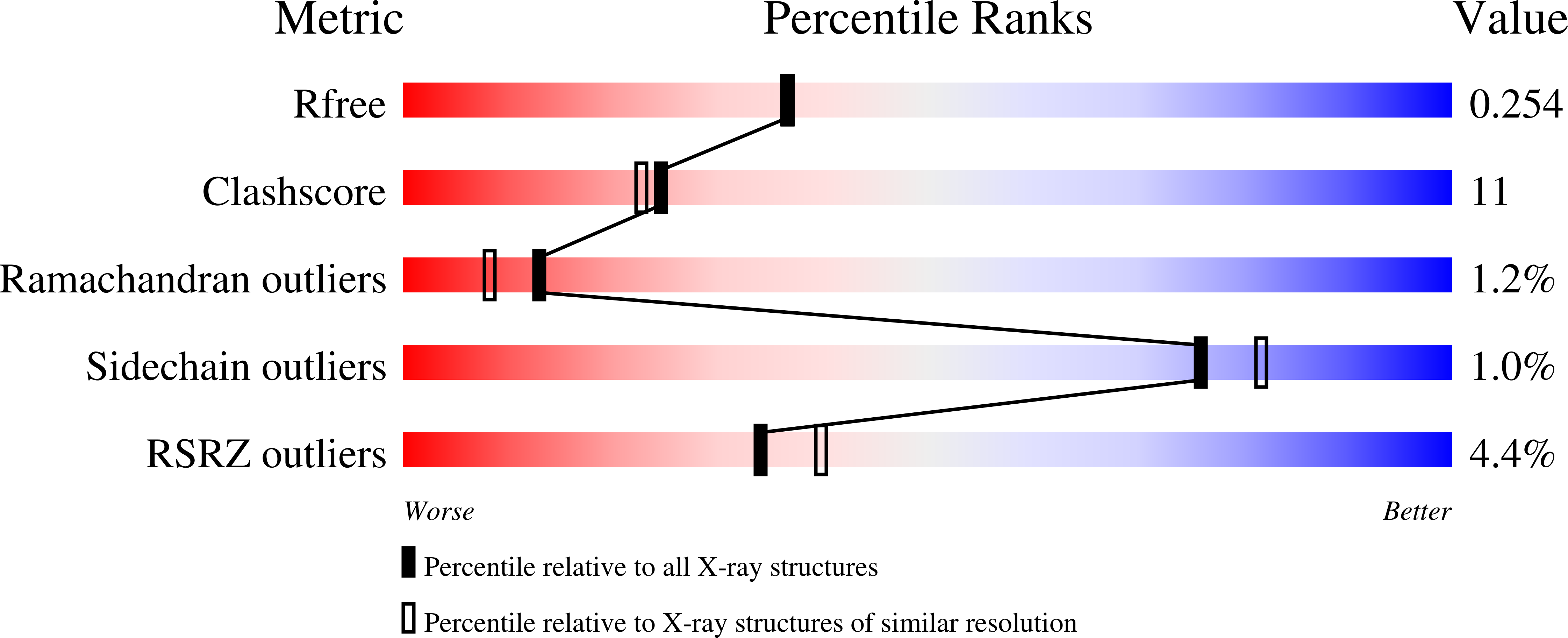
Structural Basis for Selective Recognition of Pneumococcal Cell Wall by Modular Endolysin from Phage Cp-1
Hermoso, J.A., Monterroso, B., Albert, A., Galan, B., Ahrazem, O., Garcia, P., Martinez-Ripoll, M., Garcia, J.L., Menendez, M.(2003) Structure 11: 1239
- PubMed: 14527392
- DOI: https://doi.org/10.1016/j.str.2003.09.005
- Primary Citation of Related Structures:
1H09, 1OBA - PubMed Abstract:
Pneumococcal bacteriophage-encoded lysins are modular choline binding proteins that have been shown to act as enzymatic antimicrobial agents (enzybiotics) against streptococcal infections. Here we present the crystal structures of the free and choline bound states of the Cpl-1 lysin, encoded by the pneumococcal phage Cp-1. While the catalytic module displays an irregular (beta/alpha)(5)beta(3) barrel, the cell wall-anchoring module is formed by six similar choline binding repeats (ChBrs), arranged into two different structural regions: a left-handed superhelical domain configuring two choline binding sites, and a beta sheet domain that contributes in bringing together the whole structure. Crystallographic and site-directed mutagenesis studies allow us to propose a general catalytic mechanism for the whole glycoside hydrolase family 25. Our work provides the first complete structure of a member of the large family of choline binding proteins and reveals that ChBrs are versatile elements able to tune the evolution and specificity of the pneumococcal surface proteins.
Organizational Affiliation:
Grupo de Cristalografía Macromolecular y Biología Estructural, de Macromoléculas Biológicas, Instituto Química-Física Rocasolano, CSIC, Serrano 119, 28006 Madrid, Spain. [email protected]


