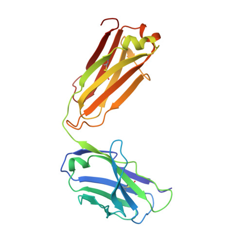
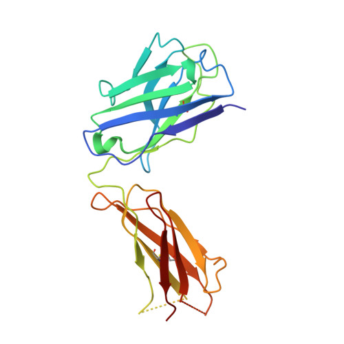
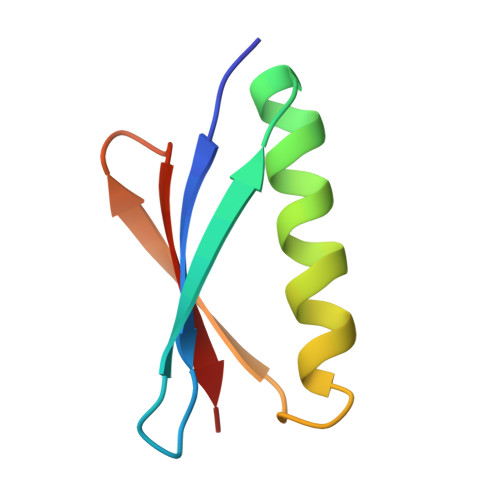
1HEZ

Structure of P. magnus protein L bound to a human IgM Fab.


Experimental Data Snapshot

  • Method: X-RAY DIFFRACTION
  • Resolution: 2.70 Å
  • R-Value Free: 0.278 
  • R-Value Work: 0.215 
  • R-Value Observed: 0.215 

Starting Model: experimental
View more details

wwPDB Validation   3D Report Full Report


This is version 1.6 of the entry. See complete history


Literature

Complex between Peptostreptococcus Magnus Protein L and a Human Antibody Reveals Structural Convergence in the Interaction Modes of Fab Binding Modes

Graille, M.Stura, E.A.Housden, N.G.Beckingham, J.A.Bottomley, S.P.Beale, D.Taussig, M.J.Sutton, B.J.Gore, M.G.Charbonnier, J.B.

(2001) Structure 9: 679

  • DOI: https://doi.org/10.1016/s0969-2126(01)00630-x
  • Primary Citation of Related Structures:  
    1HEZ

  • PubMed Abstract: 

    Peptostreptococcus magnus protein L (PpL) is a multidomain, bacterial surface protein whose presence correlates with virulence. It consists of up to five homologous immunoglobulin binding domains that interact with the variable (VL) regions of kappa light chains found on two thirds of mammalian antibodies. We refined the crystal structure of the complex between a human antibody Fab fragment (2A2) and a single PpL domain (61 residues) to 2.7 A. The asymmetric unit contains two Fab molecules sandwiching a single PpL domain, which contacts similar VL framework regions of two light chains via independent interfaces. The residues contacted on VL are remote from the hypervariable loops. One PpL-Vkappa interface agrees with previous biochemical data, while the second is novel. Site-directed mutagenesis and analytical-centrifugation studies suggest that the two PpL binding sites have markedly different affinities for VL. The PpL residues in both interactions are well conserved among different Peptostreptococcus magnus strains. The Fab contact positions identified in the complex explain the high specificity of PpL for antibodies with kappa rather than lambda chains. The PpL-Fab complex shows the first interaction of a bacterial virulence factor with a Fab light chain outside the conventional combining site. Structural comparison with two other bacterial proteins interacting with the Fab heavy chain shows that PpL, structurally homologous to streptococcal SpG domains, shares with the latter a similar binding mode. These two bacterial surface proteins interact with their respective immunoglobulin regions through a similar beta zipper interaction.


  • Organizational Affiliation

    Département d'Ingénierie et d'Etudes des Protéines, Commissariat à l'Energie Atomique, Centre d'Etudes Saclay, F-91191, Gif-sur-Yvette, France.


Macromolecules
Find similar proteins by:  (by identity cutoff)  |  3D Structure
Entity ID: 1
MoleculeChains Sequence LengthOrganismDetailsImage
KAPPA LIGHT CHAIN OF IG
A, C
214Homo sapiensMutation(s): 0 
UniProt & NIH Common Fund Data Resources
Find proteins for P01834 (Homo sapiens)
Explore P01834 
Go to UniProtKB:  P01834
PHAROS:  P01834
Entity Groups  
Sequence Clusters30% Identity50% Identity70% Identity90% Identity95% Identity100% Identity
UniProt GroupP01834
Sequence Annotations
Expand
  • Reference Sequence
Find similar proteins by:  (by identity cutoff)  |  3D Structure
Entity ID: 2
MoleculeChains Sequence LengthOrganismDetailsImage
HEAVY CHAIN OF IG
B, D
224Homo sapiensMutation(s): 0 
UniProt & NIH Common Fund Data Resources
Find proteins for P01871 (Homo sapiens)
Explore P01871 
Go to UniProtKB:  P01871
PHAROS:  P01871
GTEx:  ENSG00000211899 
Entity Groups  
Sequence Clusters30% Identity50% Identity70% Identity90% Identity95% Identity100% Identity
UniProt GroupP01871
Sequence Annotations
Expand
  • Reference Sequence
Find similar proteins by:  (by identity cutoff)  |  3D Structure
Entity ID: 3
MoleculeChains Sequence LengthOrganismDetailsImage
PROTEIN L61Finegoldia magnaMutation(s): 0 
UniProt
Find proteins for Q51918 (Finegoldia magna)
Explore Q51918 
Go to UniProtKB:  Q51918
Entity Groups  
Sequence Clusters30% Identity50% Identity70% Identity90% Identity95% Identity100% Identity
UniProt GroupQ51918
Sequence Annotations
Expand
  • Reference Sequence
Experimental Data & Validation

Experimental Data

  • Method: X-RAY DIFFRACTION
  • Resolution: 2.70 Å
  • R-Value Free: 0.278 
  • R-Value Work: 0.215 
  • R-Value Observed: 0.215 
  • Space Group: P 21 21 21
Unit Cell:
Length ( Å )Angle ( ˚ )
a = 55.185α = 90
b = 87.335β = 90
c = 210.538γ = 90
Software Package:
Software NamePurpose
CNSrefinement
HKLdata reduction
SCALEPACKdata scaling
AMoREphasing

Structure Validation

View Full Validation Report


Entry History 

Deposition Data

Revision History  (Full details and data files)

  • Version 1.0: 2001-08-10
    Type: Initial release
  • Version 1.1: 2011-05-08
    Changes: Version format compliance
  • Version 1.2: 2011-07-13
    Changes: Version format compliance
  • Version 1.3: 2018-04-18
    Changes: Data collection, Database references
  • Version 1.4: 2021-03-17
    Changes: Derived calculations, Other, Structure summary
  • Version 1.5: 2023-12-13
    Changes: Data collection, Database references, Refinement description
  • Version 1.6: 2024-11-20
    Changes: Structure summary