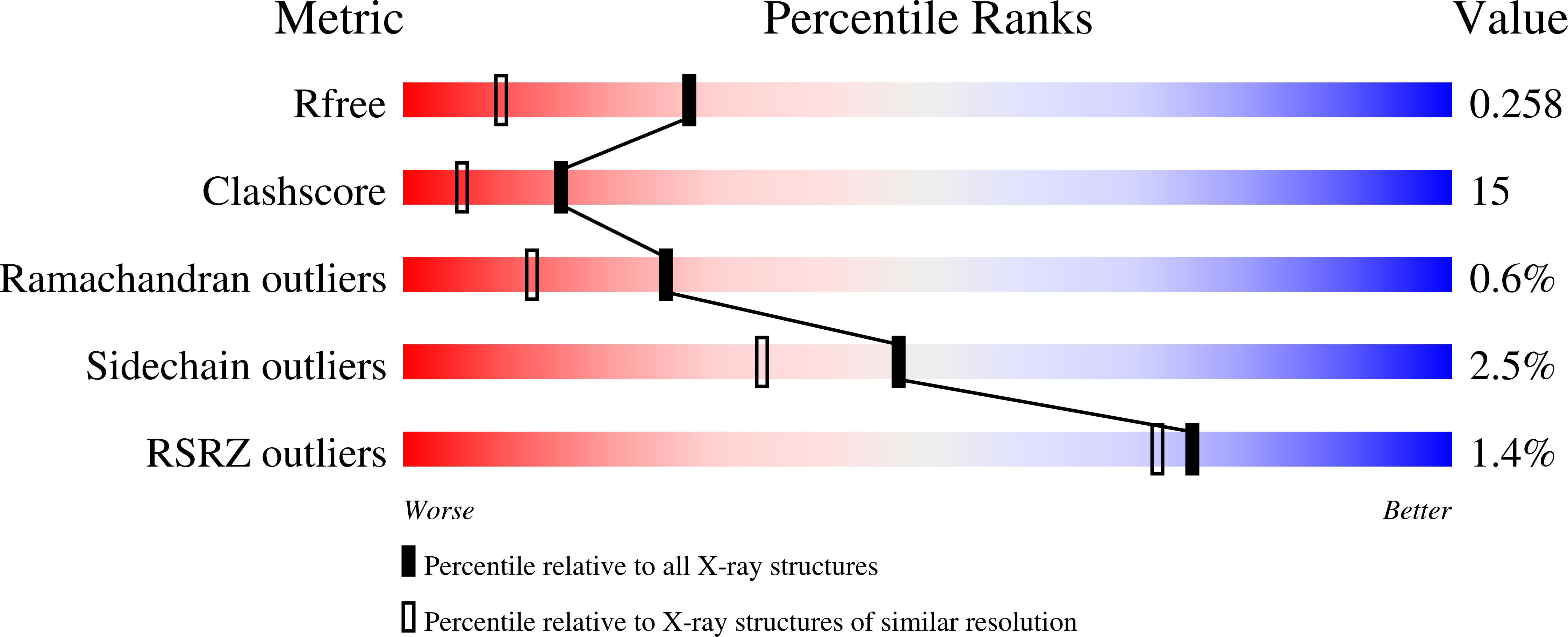
Crystal structures of the semireduced and inhibitor-bound forms of cyclic nucleotide phosphodiesterase from Arabidopsis thaliana.
Hofmann, A., Grella, M., Botos, I., Filipowicz, W., Wlodawer, A.(2002) J Biol Chem 277: 1419-1425
- PubMed: 11694509
- DOI: https://doi.org/10.1074/jbc.M107889200
- Primary Citation of Related Structures:
1JH6, 1JH7 - PubMed Abstract:
The crystal structure of the semireduced form of cyclic nucleotide phosphodiesterase (CPDase) from Arabidopsis thaliana has been solved by molecular replacement and refined at the resolution of 1.8 A. We have previously reported the crystal structure of the native form of this enzyme, whose main target is ADP-ribose 1",2"-cyclic phosphate, a product of the tRNA splicing reaction. CPDase possesses six cysteine residues, four of which are involved in forming two intra-molecular disulfide bridges. One of these bridges, between Cys-104 and Cys-110, is opened in the semireduced CPDase, whereas the other remains intact. This change of the redox state leads to a conformational rearrangement in the loop covering the active site of the protein. While the native structure shows this partially disordered loop in a coil conformation, in the semireduced enzyme the N-terminal lobe of this loop winds up and elongates the preceding alpha-helix. The semireduced state of CPDase also enabled co-crystallization with a putative inhibitor of its enzymatic activity, 2',3'-cyclic uridine vanadate. The ligand is bound within the active site, and the mode of binding is in agreement with the previously proposed enzymatic mechanism. Selected biophysical properties of the oxidized and the semireduced CPDase are also discussed.
Organizational Affiliation:
Macromolecular Crystallography Laboratory, NCI, National Institutes of Health, Frederick, Maryland 21702, USA. [email protected]


