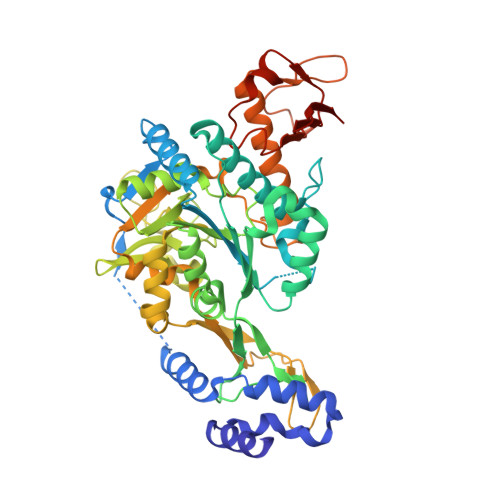
Crystal structures of two human pyrophosphorylase isoforms in complexes with UDPGlc(Gal)NAc: role of the alternatively spliced insert in the enzyme oligomeric assembly and active site architecture.
Peneff, C., Ferrari, P., Charrier, V., Taburet, Y., Monnier, C., Zamboni, V., Winter, J., Harnois, M., Fassy, F., Bourne, Y.(2001) EMBO J 20: 6191-6202
- PubMed: 11707391
- DOI: https://doi.org/10.1093/emboj/20.22.6191
- Primary Citation of Related Structures:
1JV1, 1JV3, 1JVD, 1JVG - PubMed Abstract:
The recently published human genome with its relatively modest number of genes has highlighted the importance of post-transcriptional and post-translational modifications, such as alternative splicing or glycosylation, in generating the complexities of human biology. The human UDP-N-acetylglucosamine (UDPGlcNAc) pyrophosphorylases AGX1 and AGX2, which differ in sequence by an alternatively spliced 17 residue peptide, are key enzymes synthesizing UDPGlcNAc, an essential precursor for protein glycosylation. To better understand the catalytic mechanism of these enzymes and the role of the alternatively spliced segment, we have solved the crystal structures of AGX1 and AGX2 in complexes with UDPGlcNAc (at 1.9 and 2.4 A resolution, respectively) and UDPGalNAc (at 2.2 and 2.3 A resolution, respectively). Comparison with known structures classifies AGX1 and AGX2 as two new members of the SpsA-GnT I Core superfamily and, together with mutagenesis analysis, helps identify residues critical for catalysis. Most importantly, our combined structural and biochemical data provide evidence for a change in the oligomeric assembly accompanied by a significant modification of the active site architecture, a result suggesting that the two isoforms generated by alternative splicing may have distinct catalytic properties.
Organizational Affiliation:
AFMB, UMR 6098 CNRS, 31 chemin Joseph Aiguier, 13402 Marseille Cedex 20, France.


