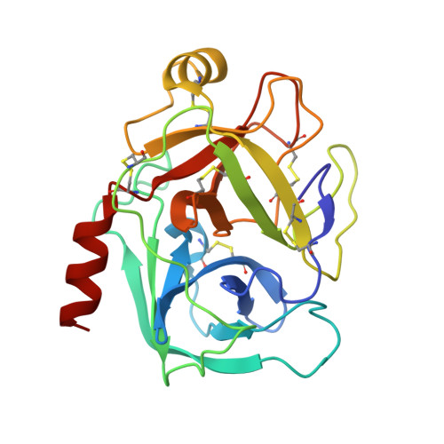
1K1N

BOVINE TRYPSIN-INHIBITOR COMPLEX


Experimental Data Snapshot

  • Method: X-RAY DIFFRACTION
  • Resolution: 2.00 Å
  • R-Value Work: 0.168 
  • R-Value Observed: 0.168 

wwPDB Validation   3D Report Full Report


Ligand Structure Quality Assessment 


This is version 1.4 of the entry. See complete history


Literature

Factorising ligand affinity: a combined thermodynamic and crystallographic study of trypsin and thrombin inhibition.

Dullweber, F.Stubbs, M.T.Musil, D.Sturzebecher, J.Klebe, G.

(2001) J Mol Biol 313: 593-614

  • DOI: https://doi.org/10.1006/jmbi.2001.5062
  • Primary Citation of Related Structures:  
    1K1I, 1K1J, 1K1L, 1K1M, 1K1N, 1K1O, 1K1P, 1K21, 1K22

  • PubMed Abstract: 

    The binding of a series of low molecular weight ligands towards trypsin and thrombin has been studied by isothermal titration calorimetry and protein crystallography. In a series of congeneric ligands, surprising changes of protonation states occur and are overlaid on the binding process. They result from induced pK(a) shifts depending on the local environment experienced by the ligand and protein functional groups in the complex (induced dielectric fit). They involve additional heat effects that must be corrected before any conclusion on the binding enthalpy (DeltaH) and entropy (DeltaS) can be drawn. After correction, trends in both contributions can be interpreted in structural terms with respect to the hydrogen bond inventory or residual ligand motions. For all inhibitors studied, a strong negative heat capacity change (DeltaC(p)) is detected, thus binding becomes more exothermic and entropically less favourable with increasing temperature. Due to a mutual compensation, Gibbs free energy remains virtually unchanged. The strong negative DeltaC(p) value cannot solely be explained by the removal of hydrophobic surface portions of the protein or ligand from water exposure. Additional contributions must be considered, presumably arising from modulations of the local water structure, changes in vibrational modes or other ordering parameters. For thrombin, smaller negative DeltaC(p) values are observed for ligand binding in the presence of sodium ions compared to the other alkali ions, probably due to stabilising effects on the protein or changes in the bound water structure.


  • Organizational Affiliation

    Philipps-Universität Marburg, Institut für Pharmazeutische Chemie, Marbacher Weg 6, D-35037 Marburg (Lahn), Germany.


Macromolecules
Find similar proteins by:  (by identity cutoff)  |  3D Structure
Entity ID: 1
MoleculeChains Sequence LengthOrganismDetailsImage
TRYPSIN223Bos taurusMutation(s): 0 
EC: 3.4.21.4
UniProt
Find proteins for P00760 (Bos taurus)
Explore P00760 
Go to UniProtKB:  P00760
Entity Groups  
Sequence Clusters30% Identity50% Identity70% Identity90% Identity95% Identity100% Identity
UniProt GroupP00760
Sequence Annotations
Expand
  • Reference Sequence
Small Molecules
Ligands 2 Unique
IDChains Name / Formula / InChI Key2D Diagram3D Interactions
CCR
Query on CCR

Download Ideal Coordinates CCD File 
C [auth A][N-[N-(4-METHOXY-2,3,6-TRIMETHYLPHENYLSULFONYL)-L-ASPARTYL]-D-(4-AMIDINO-PHENYLALANYL)]-PIPERIDINE
C29 H39 N5 O7 S
ZOXOKTJHZSUHRJ-XZOQPEGZSA-N
CA
Query on CA

Download Ideal Coordinates CCD File 
B [auth A]CALCIUM ION
Ca
BHPQYMZQTOCNFJ-UHFFFAOYSA-N
Binding Affinity Annotations 
IDSourceBinding Affinity
CCR PDBBind:  1K1N Kd: 153 (nM) from 1 assay(s)
Experimental Data & Validation

Experimental Data

  • Method: X-RAY DIFFRACTION
  • Resolution: 2.00 Å
  • R-Value Work: 0.168 
  • R-Value Observed: 0.168 
  • Space Group: P 21 21 21
Unit Cell:
Length ( Å )Angle ( ˚ )
a = 54.798α = 90
b = 58.273β = 90
c = 67.277γ = 90
Software Package:
Software NamePurpose
DENZOdata reduction
SCALEPACKdata scaling
X-PLORrefinement

Structure Validation

View Full Validation Report


Ligand Structure Quality Assessment 


Entry History 

Deposition Data

  • Released Date: 2001-11-28 
  • Deposition Author(s): Stubbs, M.T.

Revision History  (Full details and data files)

  • Version 1.0: 2001-11-28
    Type: Initial release
  • Version 1.1: 2007-10-16
    Changes: Version format compliance
  • Version 1.2: 2011-07-13
    Changes: Version format compliance
  • Version 1.3: 2012-12-12
    Changes: Other
  • Version 1.4: 2024-10-30
    Changes: Data collection, Database references, Derived calculations, Structure summary