1KSD

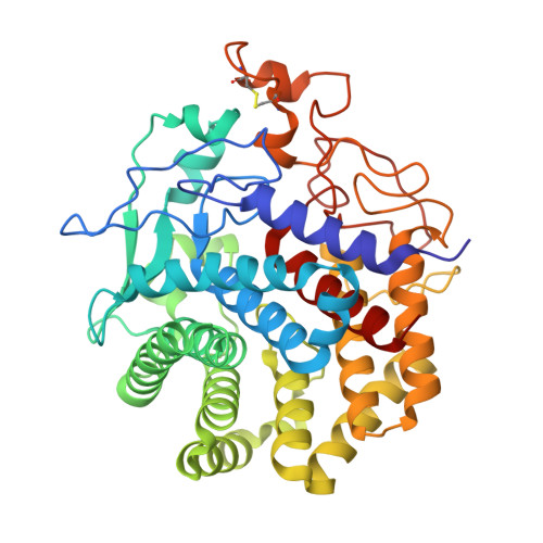
The structure of Endoglucanase from termite, Nasutitermes takasagoensis, at pH 6.5.


Experimental Data Snapshot

  • Method: X-RAY DIFFRACTION
  • Resolution: 1.60 Å
  • R-Value Free: 0.193 
  • R-Value Work: 0.170 

Starting Model: experimental
View more details

wwPDB Validation   3D Report Full Report


This is version 1.4 of the entry. See complete history


Literature

Structure of an endoglucanase from termite, Nasutitermes takasagoensis.

Khademi, S.Guarino, L.A.Watanabe, H.Tokuda, G.Meyer, E.F.

(2002) Acta Crystallogr D Biol Crystallogr 58: 653-659

  • DOI: https://doi.org/10.1107/s0907444902002366
  • Primary Citation of Related Structures:  
    1KS8, 1KSC, 1KSD

  • PubMed Abstract: 

    Contrary to conventional wisdom, it has been shown recently that termites do not necessarily depend on symbiotic bacteria to process cellulose. They secrete their own cellulases, mainly endo-beta-1,4-glucanase and beta-1,4-glucosidase. Here, the first structure of an endogenous endoglucanase from the higher termite Nasutitermes takasagoensis (NtEgl) is reported at 1.40 A resolution. NtEgl has the general folding of an (alpha/alpha)(6) barrel, which is a common folding pattern for glycosyl hydrolase family 9. Three-dimensional structural analysis shows that the conserved Glu412 is the catalytic acid/base residue and the conserved Asp54 or Asp57 is the base. The enzyme has a Ca(2+)-binding site near its substrate-binding cleft. Comparison between the structure of the Ca(2+)-free enzyme produced by reducing the pH of the soaked crystal from 5.6 (the pH of optimum enzyme activity) to 2.5 with that of the Ca(2+)-bound enzyme did not show significant differences in the locations of the C(alpha) atoms. The main differences are in the conformation of the residue side chains ligating the Ca(2+) ion. The overall structure of NtEgl at pH 6.5 is similar to that at pH 5.6. The major change observed was in the conformation of the side chain of the catalytic acid/base Glu412, which rotates from a hydrophobic cavity to a relatively hydrophilic environment. This side-chain displacement may decrease the enzyme activity at higher pH.


  • Organizational Affiliation

    Biographics Laboratory, Texas A&M University, Department of Biochemistry and Biophysics, College Station, TX 77843-2128, USA.


Macromolecules
Find similar proteins by:  (by identity cutoff)  |  3D Structure
Entity ID: 1
MoleculeChains Sequence LengthOrganismDetailsImage
Endo-b-1,4-glucanase433Nasutitermes takasagoensisMutation(s): 0 
EC: 3.2.1.4
UniProt
Find proteins for O77044 (Nasutitermes takasagoensis)
Explore O77044 
Go to UniProtKB:  O77044
Entity Groups  
Sequence Clusters30% Identity50% Identity70% Identity90% Identity95% Identity100% Identity
UniProt GroupO77044
Sequence Annotations
Expand
  • Reference Sequence
Small Molecules
Ligands 1 Unique
IDChains Name / Formula / InChI Key2D Diagram3D Interactions
CA
Query on CA

Download Ideal Coordinates CCD File 
B [auth A]CALCIUM ION
Ca
BHPQYMZQTOCNFJ-UHFFFAOYSA-N
Experimental Data & Validation

Experimental Data

  • Method: X-RAY DIFFRACTION
  • Resolution: 1.60 Å
  • R-Value Free: 0.193 
  • R-Value Work: 0.170 
  • Space Group: C 1 2 1
Unit Cell:
Length ( Å )Angle ( ˚ )
a = 89.251α = 90
b = 83.924β = 95.47
c = 60.878γ = 90
Software Package:
Software NamePurpose
DENZOdata reduction
SCALEPACKdata scaling
CNSrefinement
CNSphasing

Structure Validation

View Full Validation Report


Entry History 

Deposition Data

Revision History  (Full details and data files)

  • Version 1.0: 2003-01-21
    Type: Initial release
  • Version 1.1: 2008-04-27
    Changes: Version format compliance
  • Version 1.2: 2011-07-13
    Changes: Version format compliance
  • Version 1.3: 2023-08-16
    Changes: Data collection, Database references, Derived calculations, Refinement description
  • Version 1.4: 2024-10-16
    Changes: Structure summary