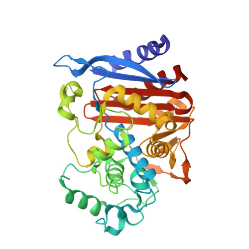
1L2S

X-ray crystal structure of AmpC beta-lactamase from E. coli in complex with a DOCK-predicted non-covalent inhibitor


Experimental Data Snapshot

  • Method: X-RAY DIFFRACTION
  • Resolution: 1.94 Å
  • R-Value Free: 0.207 
  • R-Value Work: 0.173 

Starting Model: experimental
View more details

wwPDB Validation   3D Report Full Report


Ligand Structure Quality Assessment 


This is version 1.3 of the entry. See complete history


Literature

Structure-based discovery of a novel, noncovalent inhibitor of AmpC beta-lactamase.

Powers, R.A.Morandi, F.Shoichet, B.K.

(2002) Structure 10: 1013-1023

  • DOI: https://doi.org/10.1016/s0969-2126(02)00799-2
  • Primary Citation of Related Structures:  
    1L2S

  • PubMed Abstract: 

    beta-lactamases are the most widespread resistance mechanisms to beta-lactam antibiotics, and there is a pressing need for novel, non-beta-lactam drugs. A database of over 200,000 compounds was docked to the active site of AmpC beta-lactamase to identify potential inhibitors. Fifty-six compounds were tested, and three had K(i) values of 650 microM or better. The best of these, 3-[(4-chloroanilino)sulfonyl]thiophene-2-carboxylic acid, was a competitive noncovalent inhibitor (K(i) = 26 microM), which also reversed resistance to beta-lactams in bacteria expressing AmpC. The structure of AmpC in complex with this compound was determined by X-ray crystallography to 1.94 A and reveals that the inhibitor interacts with key active-site residues in sites targeted in the docking calculation. Indeed, the experimentally determined conformation of the inhibitor closely resembles the prediction. The structure of the enzyme-inhibitor complex presents an opportunity to improve binding affinity in a novel series of inhibitors discovered by structure-based methods.


  • Organizational Affiliation

    Department of Molecular Pharmacology and Biological Chemistry, Northwestern University, 303 East Chicago Avenue, IL 60611, USA.


Macromolecules
Find similar proteins by:  (by identity cutoff)  |  3D Structure
Entity ID: 1
MoleculeChains Sequence LengthOrganismDetailsImage
beta-lactamase
A, B
358Escherichia coliMutation(s): 0 
Gene Names: K12
EC: 3.5.2.6
UniProt
Find proteins for P00811 (Escherichia coli (strain K12))
Explore P00811 
Go to UniProtKB:  P00811
Entity Groups  
Sequence Clusters30% Identity50% Identity70% Identity90% Identity95% Identity100% Identity
UniProt GroupP00811
Sequence Annotations
Expand
  • Reference Sequence
Small Molecules
Binding Affinity Annotations 
IDSourceBinding Affinity
STC BindingDB:  1L2S Ki: 2.60e+4 (nM) from 1 assay(s)
PDBBind:  1L2S Ki: 2.60e+4 (nM) from 1 assay(s)
Experimental Data & Validation

Experimental Data

  • Method: X-RAY DIFFRACTION
  • Resolution: 1.94 Å
  • R-Value Free: 0.207 
  • R-Value Work: 0.173 
  • Space Group: C 1 2 1
Unit Cell:
Length ( Å )Angle ( ˚ )
a = 118.67α = 90
b = 76.42β = 116.63
c = 97.9γ = 90
Software Package:
Software NamePurpose
CNSrefinement
DENZOdata reduction
SCALEPACKdata scaling
CNSphasing

Structure Validation

View Full Validation Report


Ligand Structure Quality Assessment 


Entry History 

Deposition Data

Revision History  (Full details and data files)

  • Version 1.0: 2002-07-17
    Type: Initial release
  • Version 1.1: 2008-04-28
    Changes: Version format compliance
  • Version 1.2: 2011-07-13
    Changes: Version format compliance
  • Version 1.3: 2023-08-16
    Changes: Data collection, Database references, Derived calculations, Refinement description