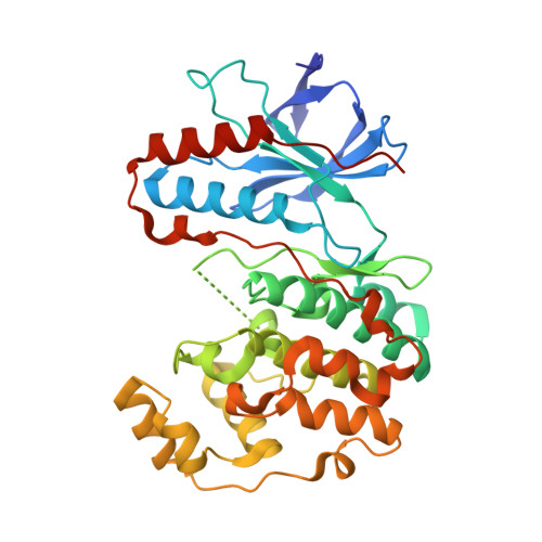
1LEW

CRYSTAL STRUCTURE OF MAP KINASE P38 COMPLEXED TO THE DOCKING SITE ON ITS NUCLEAR SUBSTRATE MEF2A


Experimental Data Snapshot

  • Method: X-RAY DIFFRACTION
  • Resolution: 2.30 Å
  • R-Value Free: 0.257 
  • R-Value Work: 0.215 
  • R-Value Observed: 0.235 

wwPDB Validation   3D Report Full Report


This is version 1.5 of the entry. See complete history


Literature

Crystal structures of MAP kinase p38 complexed to the docking sites on its nuclear substrate MEF2A and activator MKK3b.

Chang, C.I.Xu, B.E.Akella, R.Cobb, M.H.Goldsmith, E.J.

(2002) Mol Cell 9: 1241-1249

  • DOI: https://doi.org/10.1016/s1097-2765(02)00525-7
  • Primary Citation of Related Structures:  
    1LEW, 1LEZ

  • PubMed Abstract: 

    The structures of the MAP kinase p38 in complex with docking site peptides containing a phi(A)-X-phi(B) motif, derived from substrate MEF2A and activating enzyme MKK3b, have been solved. The peptides bind to the same site in the C-terminal domain of the kinase, which is both outside the active site and distinct from the "CD" domain previously implicated in docking site interactions. Mutational analysis on the interaction of p38 with the docking sites supports the crystallographic models and has uncovered two novel residues on the docking groove that are critical for binding. The two peptides induce similar large conformational changes local to the peptide binding groove. The peptides also induce unexpected and different conformational changes in the active site, as well as structural disorder in the phosphorylation lip.


  • Organizational Affiliation

    Department of Biochemistry, The University of Texas Southwestern Medical Center at Dallas, 5323 Harry Hines Boulevard, Dallas, TX 75390, USA.


Macromolecules
Find similar proteins by:  (by identity cutoff)  |  3D Structure
Entity ID: 1
MoleculeChains Sequence LengthOrganismDetailsImage
Mitogen-activated protein kinase 14360Mus musculusMutation(s): 0 
Gene Names: Mapk14Crk1Csbp1Csbp2
EC: 2.7.11.24
UniProt
Find proteins for P47811 (Mus musculus)
Explore P47811 
Go to UniProtKB:  P47811
Entity Groups  
Sequence Clusters30% Identity50% Identity70% Identity90% Identity95% Identity100% Identity
UniProt GroupP47811
Sequence Annotations
Expand
  • Reference Sequence

Find similar proteins by:  Sequence   |   3D Structure  

Entity ID: 2
MoleculeChains Sequence LengthOrganismDetailsImage
Myocyte-specific enhancer factor 2A12Homo sapiensMutation(s): 0 
UniProt & NIH Common Fund Data Resources
Find proteins for Q02078 (Homo sapiens)
Explore Q02078 
Go to UniProtKB:  Q02078
PHAROS:  Q02078
GTEx:  ENSG00000068305 
Entity Groups  
Sequence Clusters30% Identity50% Identity70% Identity90% Identity95% Identity100% Identity
UniProt GroupQ02078
Sequence Annotations
Expand
  • Reference Sequence
Experimental Data & Validation

Experimental Data

  • Method: X-RAY DIFFRACTION
  • Resolution: 2.30 Å
  • R-Value Free: 0.257 
  • R-Value Work: 0.215 
  • R-Value Observed: 0.235 
  • Space Group: P 31 2 1
Unit Cell:
Length ( Å )Angle ( ˚ )
a = 81.873α = 90
b = 81.873β = 90
c = 123.016γ = 120
Software Package:
Software NamePurpose
CNSrefinement
DENZOdata reduction
SCALEPACKdata scaling
CNSphasing

Structure Validation

View Full Validation Report


Entry History 

Deposition Data

Revision History  (Full details and data files)

  • Version 1.0: 2002-07-10
    Type: Initial release
  • Version 1.1: 2008-04-28
    Changes: Version format compliance
  • Version 1.2: 2011-07-13
    Changes: Version format compliance
  • Version 1.3: 2012-02-15
    Changes: Database references
  • Version 1.4: 2017-06-21
    Changes: Database references, Source and taxonomy, Structure summary
  • Version 1.5: 2024-02-14
    Changes: Data collection, Database references