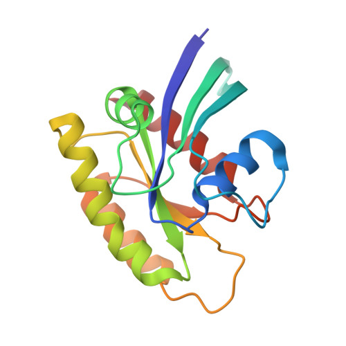
1LF5

Crystal Structure of RasA59G in the GDP-bound Form


Experimental Data Snapshot

  • Method: X-RAY DIFFRACTION
  • Resolution: 1.70 Å
  • R-Value Free: 0.245 
  • R-Value Work: 0.222 

Starting Model: experimental
View more details

wwPDB Validation   3D Report Full Report


This is version 2.1 of the entry. See complete history


Literature

The Structural Basis for the Transition from Ras-GTP to Ras-GDP

Hall, B.E.Bar-Sagi, D.Nassar, N.

(2002) Proc Natl Acad Sci U S A 99: 12138-12142

  • DOI: https://doi.org/10.1073/pnas.192453199
  • Primary Citation of Related Structures:  
    1LF0, 1LF5

  • PubMed Abstract: 

    The conformational changes in Ras that accompany the hydrolysis of GTP are critical to its function as a molecular switch in signaling pathways. Understanding how GTP is hydrolyzed by revealing the sequence of intermediary structures in the reaction is essential for understanding Ras signaling. Until now, no structure of an intermediate in GTP hydrolysis has been experimentally determined for Ras alone. We have solved the crystal structure of the Ala-59 to Gly mutant of Ras, (RasA59G), bound to guanosine 5'-imidotriphosphate or GDP to 1.7-A resolution. In the guanosine 5'-imidotriphosphate-bound form, this mutant adopts a conformation that is intermediate between the GTP- and GDP-bound forms of wild-type Ras and that is similar to what has been predicted by molecular dynamics simulation [Ma, J. P. & Karplus, M. (1997) Proc. Natl. Acad. Sci. USA 94, 11905-11910]. This conformation is stabilized by direct and water-mediated interactions between the switch 1 and switch 2 regions and is characterized by an increase in the binding affinity for GTP. We propose that the structural changes promoted by the Ala-59 to Gly mutation exhibit a discrete conformational state assumed by wild-type Ras during GTP hydrolysis.


  • Organizational Affiliation

    Graduate Program in Molecular and Cellular Pharmacology, Department of Molecular Genetics, Stony Brook University, Stony Brook, NY 11794-8661, USA.


Macromolecules
Find similar proteins by:  (by identity cutoff)  |  3D Structure
Entity ID: 1
MoleculeChains Sequence LengthOrganismDetailsImage
Transforming protein P21/H-RAS-1166Homo sapiensMutation(s): 1 
EC: 3.6.5.2
UniProt & NIH Common Fund Data Resources
Find proteins for P01112 (Homo sapiens)
Explore P01112 
Go to UniProtKB:  P01112
PHAROS:  P01112
GTEx:  ENSG00000174775 
Entity Groups  
Sequence Clusters30% Identity50% Identity70% Identity90% Identity95% Identity100% Identity
UniProt GroupP01112
Sequence Annotations
Expand
  • Reference Sequence
Small Molecules
Ligands 2 Unique
IDChains Name / Formula / InChI Key2D Diagram3D Interactions
GDP
Query on GDP

Download Ideal Coordinates CCD File 
C [auth A]GUANOSINE-5'-DIPHOSPHATE
C10 H15 N5 O11 P2
QGWNDRXFNXRZMB-UUOKFMHZSA-N
MG
Query on MG

Download Ideal Coordinates CCD File 
B [auth A]MAGNESIUM ION
Mg
JLVVSXFLKOJNIY-UHFFFAOYSA-N
Experimental Data & Validation

Experimental Data

  • Method: X-RAY DIFFRACTION
  • Resolution: 1.70 Å
  • R-Value Free: 0.245 
  • R-Value Work: 0.222 
  • Space Group: H 3 2
Unit Cell:
Length ( Å )Angle ( ˚ )
a = 93.112α = 90
b = 93.112β = 90
c = 120.301γ = 120
Software Package:
Software NamePurpose
DENZOdata reduction
SCALEPACKdata scaling
AMoREphasing
CNSrefinement

Structure Validation

View Full Validation Report


Entry History 

Deposition Data

Revision History  (Full details and data files)

  • Version 1.0: 2002-11-06
    Type: Initial release
  • Version 1.1: 2008-04-28
    Changes: Version format compliance
  • Version 1.2: 2011-07-13
    Changes: Version format compliance
  • Version 1.3: 2011-11-16
    Changes: Atomic model
  • Version 2.0: 2021-10-27
    Changes: Atomic model, Database references, Derived calculations
  • Version 2.1: 2023-08-16
    Changes: Advisory, Data collection, Derived calculations, Refinement description