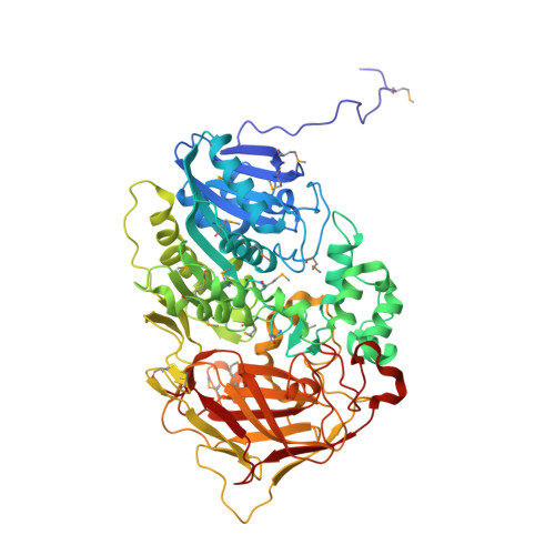
1MPX

ALPHA-AMINO ACID ESTER HYDROLASE LABELED WITH SELENOMETHIONINE


Experimental Data Snapshot

  • Method: X-RAY DIFFRACTION
  • Resolution: 1.90 Å
  • R-Value Free: 0.178 
  • R-Value Work: 0.149 
  • R-Value Observed: 0.150 

wwPDB Validation   3D Report Full Report


This is version 1.4 of the entry. See complete history


Literature

The sequence and crystal structure of the alpha-amino acid ester hydrolase from Xanthomonas citri define a new family of beta-lactam antibiotic acylases.

Barends, T.R.Polderman-Tijmes, J.J.Jekel, P.A.Hensgens, C.M.de Vries, E.J.Janssen, D.B.Dijkstra, B.W.

(2003) J Biol Chem 278: 23076-23084

  • DOI: https://doi.org/10.1074/jbc.M302246200
  • Primary Citation of Related Structures:  
    1MPX

  • PubMed Abstract: 

    alpha-Amino acid ester hydrolases (AEHs) catalyze the hydrolysis and synthesis of esters and amides with an alpha-amino group. As such, they can synthesize beta-lactam antibiotics from acyl compounds and beta-lactam nuclei obtained from the hydrolysis of natural antibiotics. This article describes the gene sequence and the 1.9-A resolution crystal structure of the AEH from Xanthomonas citri. The enzyme consists of an alpha/beta-hydrolase fold domain, a helical cap domain, and a jellyroll beta-domain. Structural homology was observed to the Rhodococcus cocaine esterase, indicating that both enzymes belong to the same class of bacterial hydrolases. Docking of a beta-lactam antibiotic in the active site explains the substrate specificity, specifically the necessity of an alpha-amino group on the substrate, and explains the low specificity toward the beta-lactam nucleus.


  • Organizational Affiliation

    Department of Biophysical Chemistry, Groningen Biomolecular Sciences and Biotechnology Institute, University of Groningen, Nijenborgh 4, NL-9747 AG Groningen, The Netherlands.


Macromolecules
Find similar proteins by:  (by identity cutoff)  |  3D Structure
Entity ID: 1
MoleculeChains Sequence LengthOrganismDetailsImage
alpha-amino acid ester hydrolase
A, B, C, D
615Xanthomonas citriMutation(s): 18 
EC: 3.1.1.43
UniProt
Find proteins for Q6YBS3 (Xanthomonas citri)
Explore Q6YBS3 
Go to UniProtKB:  Q6YBS3
Entity Groups  
Sequence Clusters30% Identity50% Identity70% Identity90% Identity95% Identity100% Identity
UniProt GroupQ6YBS3
Sequence Annotations
Expand
  • Reference Sequence
Small Molecules
Ligands 2 Unique
IDChains Name / Formula / InChI Key2D Diagram3D Interactions
GOL
Query on GOL

Download Ideal Coordinates CCD File 
F [auth A]
G [auth A]
H [auth A]
J [auth B]
K [auth B]
F [auth A],
G [auth A],
H [auth A],
J [auth B],
K [auth B],
L [auth B],
N [auth C],
O [auth C],
Q [auth D],
R [auth D]
GLYCEROL
C3 H8 O3
PEDCQBHIVMGVHV-UHFFFAOYSA-N
CA
Query on CA

Download Ideal Coordinates CCD File 
E [auth A],
I [auth B],
M [auth C],
P [auth D]
CALCIUM ION
Ca
BHPQYMZQTOCNFJ-UHFFFAOYSA-N
Modified Residues  1 Unique
IDChains TypeFormula2D DiagramParent
MSE
Query on MSE
A, B, C, D
L-PEPTIDE LINKINGC5 H11 N O2 SeMET
Experimental Data & Validation

Experimental Data

  • Method: X-RAY DIFFRACTION
  • Resolution: 1.90 Å
  • R-Value Free: 0.178 
  • R-Value Work: 0.149 
  • R-Value Observed: 0.150 
  • Space Group: P 1 21 1
Unit Cell:
Length ( Å )Angle ( ˚ )
a = 89.772α = 90
b = 126.016β = 91.06
c = 132.291γ = 90
Software Package:
Software NamePurpose
DENZOdata reduction
SCALEPACKdata scaling
SnBphasing
SOLVEphasing
RESOLVEmodel building
ARP/wARPmodel building
REFMACrefinement
RESOLVEphasing

Structure Validation

View Full Validation Report


Entry History 

Deposition Data

Revision History  (Full details and data files)

  • Version 1.0: 2003-04-15
    Type: Initial release
  • Version 1.1: 2008-04-28
    Changes: Version format compliance
  • Version 1.2: 2011-07-13
    Changes: Non-polymer description, Version format compliance
  • Version 1.3: 2017-10-11
    Changes: Refinement description
  • Version 1.4: 2024-10-30
    Changes: Data collection, Database references, Derived calculations, Structure summary